PDF hosted at the Radboud Repository of the Radboud University
Nijmegen
The following full text is a publisher's version.
For additional information about this publication click this link.
http://hdl.handle.net/2066/23088
Please be advised that this information was generated on 2015-01-28 and may be subject to
change.
JOURNAL OF PATHOLOGY, VOL. 1 7 9 : 2 6 0 -2 6 5 (1 9 9 6 )
TETRANECTIN AND PLASMIN/PLASMINOGEN ARE
SIMILARLY DISTRIBUTED AT THE INVASIVE FRONT
OF CUTANEOUS MELANOMA LESIONS
TEUNIS J. DE VRIES, PETER E. J. D E W IT , IN G E CLEM M ENSEN*, H E IN W . V E R S P A G E T t, U L R IC H I-I. W E ID L E f,
EVA B, BRO CK ER§, D IR K J. RUTTER A N D GOOS N , P . VAN MUIJEN
Department o f Pathology, University Hospital\ Nijmegen, The Netherlands; *DAKO AIS, Glostrup, Denmark; |Department o f
Gastroenterology and Hepatology , University Hospital , Leiden, The Netherlands; \Boehringer Mannheim , Penzberg, Germany;
§,Department o f Dermatology, University Hospital Wurzburg, Germany
SUMMARY
The induction of expression of the components of the proteolytic plasminogen activation system in cutaneous melanocytic tumour
progression has previously been reported. Plasminogen activators, their inhibitors, and the receptor for urokinase were present only in
advanced primary melanomas and melanoma metastases. The present study reports on the presence of tetranectin and plasmin/
plasminogen, two proteins connected with plasminogen activation, in cutaneous melanocytic lesions. The distribution of tetranectin and
plasminogen was studied by immunohistochemistry in 105 freshly frozen melanocytic lesions of common naevocellular naevi («=24),
atypical naevi (/i=14), early (« = 12) and advanced («=20) primary melanomas, and melanoma metastases («=35). Both tetrancctin and
plasminogen were detected in a variety of tissue components. In all stages of melanocytic tumour progression, tetranectin was found in
endothelium, perivascular dendritic cells, and leukocytes. Plasminogen was present in endothelium and in the basal layer of the normal
skin. Tetranectin and plasminogen staining of fibroblastic cells at the invasive front and of extracellular matrix was, however, restricted
to malignant lesions. Co-localization of tetranectin and plasminogen was found in 50 per cent of the early primary melanomas and in
more than 75 per cent of the advanced melanomas and melanoma metastases. These results suggest a coordinated role for tetranectin
and plasminogen at the invasive front of melanomas. Tetranectin-bound plasminogen may facilitate the migration of tumour cells.
KEY w o r d s — melanoma;
skin; tetranectin; plasminogen; plasminogen activation; immunohistochemistry; co-localization
INTRODUCTION
In the concept o f tum our cell invasion and metastasis,
the degradation o f tissue barriers has been implicated, In
this process, several proteolytic enzyme systems can be
recruited ; 1*2 one system involved in tum our spread is
plasminogen activation . 3*4 In vitro5 and in vivo6'* find
ings indicate th at both tu m o u r cells and fibroblastic
stromal cells are involved in the plasminogen activation
process at the tu m o u r-stro m a interface . 9 In cutaneous
melanoma lesions, urokinase plasminogen activator
(u-PA) in particular is m arkedly present in tum our
cells8 J 0 and in stromal cells 8 at the periphery o f tum our
nodules.
Two other molecules which are involved in the process
o f plasminogen activation and which play a putative role
in the migration process o f tu m o u r cells are plasminogen
and tetranectin. Tetranectin is a 6 8 kD protein purified
from plasma, which binds to the kringle 4 domain of
plasminogen . 13 Plasma o f cancer patients contains sig
nificantly lower levels o f tetranectin com pared to ageand sex-matched control groups . 12-14 Tetranectin is
present in a variety of cells, including monocytes , 15
At the time of submission o f this manuscript, Ms I. Clemmensen
passed away due to the disease she knew so well. As the original
discoverer of tetranectin, she contributed on the role of tetranectin in
various neoplasms. This article is dedicated to her scientific work.
Addressee for correspondence: Teun de Vries, Department of
Pathology, University Hospital, P.O. Box 9101, 6500 HB Nijmegen,
The Netherlands.
C CC 0 0 2 2 -3 4 1 7 /9 6 /0 7 0 2 6 0 -0 6
© 1996 by Joh n W iley & S o n s, Ltd.
neutrophils , 16 and fibroblasts . 17 In breast , 18 ovarian , 14
and colonic 19,20 tumours, tetranectin is present in the
strom a surrounding the tum our. Moreover, plasma
tetranectin content and stromal immunoreactivity are of
prognostic relevance in patients with ovarian cancer . 14
Plasminogen is the substrate for plasminogen acti
vators. Cleavage o f plasminogen by plasminogen
activators form s the active enzyme plasmin. Little is
known ab o u t the presence of plasmin/plasminogen in
cancerous tissues. In colonic carcinomas, it is reported
to be present in stromal cells .21
Recently, we described the presence of the compo
nents of the plasminogen activation system in advanced
stages o f hu m an cutaneous melanocytic tumour progres
sion .8 Here, we present data on the distribution of
tetranectin and plasminogen in cutaneous melanocytic
lesions.
MATERIALS AND METHODS
Tissue specimens
Representative tissue samples were freshly received
from cutaneous melanocytic lesions excised from
patients at the University Hospital, Nijmegen, The
Netherlands, and from patients at the University
Hospital, W urzburg, Germany. They were snap-frozen
in liquid nitrogen and stored at — 80°C until sectioning.
Based on conventional histopathological examination of
paraffin sections, lesions were divided into five classes:
common naevocellular naevus (NN, n= 24), atypical
Received 29 May 1995
Accepted 8 December 1995
TETRANECTIN AND PLASMINOGEN IN MELANOMA
naevus (AN, n - 14), early primary melanoma (Breslow
thickness <1*5 mm) (ePM, 77= 12), advanced prim ary
m elanom a (Breslow thickness >1-5 mm) (aPM , «=20),
an d m elanom a metastasis (M M , n = 35).
261
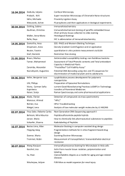
metastases were stained for tetranectin and p lasm in o
gen. Typical examples o f tetranectin a n d plasm inogen
staining are shown in Figs 1 and 2 .
Tetranectin
Antibodies
T w o well-characterized antibodies were used. R abbit
polyclonal antibody A371 against tetranectin11 has been
used previously in immunohistochemistry. Staining
w ith this polyclonal antibody compared well to the
staining obtained with a monoclonal antibody against
tetran ectin . 18’22
R a b b it polyclonal antibody against plasminogen/
plasm in was affinity-purified by passaging through a
Sepharose column to which the kringle 1-3 p a rt o f
plasm inogen was coupled. Others have show n that
immunohistochemical staining of tissue sections with
this antibody compares well to various m onoclonal
a n d polyclonal antibodies against plasmin/plasminogen
(C hristensen et al., paper submitted).
Immunohis to chemis try
T h e immunohistochemical staining procedure for
polyclonal antibodies was as described previously . 8
Briefly, 4 fi m cryostat sections were air-dried overnight
a t ro o m temperature and stored at —80°C until use.
A fte r acetone fixation, dilutions o f the polyclonal anti
bodies were applied to the sections. A ntibody bind
ing was visualized with a peroxidase-labelled swine
a n ti-ra b b it secondary antibody and incubation with
3-amino-9-ethylcarbazole as a substrate for peroxidase.
A fter counterstaining the nuclei with P apanicolaou’s
H a rris solution, sections were mounted with K aiser’s
glycerin (Merck, D arm stadt, Germany). An incubation
in which the first antibody was omitted served as a
negative control. Consecutive sections of the same lesion
w ere incubated with the tetranectin and plasminogen
antibodies. Localization similarities were judged by
c o m p a rin g the staining results of the two sections.
Score
F o r each section, the percentage of positive melanocytic cells was estimated. Each section was assigned to
one o f the following categories: 0 per cent, 1-5 per cent,
6-25 per cent, and 26-100 per cent positivity. Positive
m elanocytic staining was registered when at least
I per cent of the melanocytic cells stained. N otes were
taken o f other staining components (fibroblast-like cells,
extracellular matrix) am ong the melanocytic areas.
Sections were scored independently by two observers
(T, J. de V., P. E. J. de W.). Discrepancies were found in
fewer th an 10 per cent o f the lesions. These cases were
re-evaluated jointly until agreement was reached.
RESULTS
Tissue sections of naevocellular naevi, atypical naevi,
early a n d advanced prim ary melanomas, and m elanom a
The various aspects o f tetranectin staining are su m
marized in Table I. Tetranectin was localized in a variety
o f cell types. In benign com m on naevocellular naevi a n d
in atypical naevi, there were scattered positively stained
cells in the dermis, so-called perivascular dendritic cells 21
(Fig. la). In addition, some blood vessels showed p o si
tive endothelium (not shown). Tetranectin-positive
cells were often found in the lum en of blo od vessels, in
both benign and m alignant lesions (Fig. lb). Staining o f
the extracellular m atrix surrounding tu m o u r cells w as
observed in prim ary m elanom as and m etastases only.
Fibroblastic cells either did not stain, o r stained weakly
in benign lesions, Staining of the stro m a was frequently
observed in m alignant lesions an d was m u ch m ore
pronounced, especially a t the invasive fro n t (Fig. Ik).
N o tetranectin staining o f melanocytic cells was encoun
tered in the benign lesions and tetranectin staining o f
tu m o u r cells was seen in very few m align an t lesions
(Fig. 2a). Staining m ainly in the 1-5 per cent category
was found in 1 / 1 2 o f the early prim ary tum ours, in 1 / 2 0
o f the advanced prim ary m elanom as and in 4/35 o f the
m etastatic lesions. O ne advanced prim ary m elan o m a
contained 6-25 per cent tetranectin-positive cells.
Plasminogen
T he various aspects o f plasm inogen staining are su m
marized in Table II. Plasminogen was found in the basal
cell layer of the epidermis, b o th in benign and in
m alignant lesions (Fig. lc). This staining was often m o re
pronounced tow ards the basal m em brane (not shown).
In all types o f lesions studied, there was staining of the
endothelium o f blood vessels (not shown). T h e ex tra
cellular m atrix around tum our cells stained in m a n y
advanced prim ary m elanom as and in various m etastases
(Fig. le). Staining of extracellular m atrix was m ore often
seen, and m ore intense th an in the case o f tetranectin.
N o staining or weak staining o f fibroblastic cells w as
encountered in benign lesions an d stronger staining o f
the stroma, m arkedly at the tu m o u r-s tro m a interface
(Figs li and lj), was found in prim ary m elan o m a and in
m elanom a metastases.
Plasminogen staining o f melanocytic cells was absent
in the com m on naevocellular naevi and in the atypical
naevi, whereas some o f the m alignant lesions (Fig. 2b)
showed positivity. T u m o u r cell staining was encountered
in only a few cases of prim ary m elano m a and m elan o m a
metastases. This staining, mainly in the 1-5 per cent
category, was present in 0 / 1 2 o f the early prim ary
tum ours, in 1 / 2 0 o f the advanced p rim a ry m elanom as
a n d in 7/35 o f the m etastatic lesions. One advanced
prim ary m elanom a and one m etastasis contained
6-25 per cent plasminogen-positive cells.
Co-localization o f tetranectin and plasminogen
In the m ajority of m alignant lesions, tetranectin a n d
plasminogen were found in corresponding cell types o r
Wi0$
illlS iS S iiii
é$ÄB
iip
ita
»
fe;5%
■il „.„iiSIpsIliïlif
i/.'.y .y - ; : ," ,',
& . V ,Y
: : i V | r ::>''
11
iiv v /
I
■ y .-:
VA
I'-/-;
ü ir ^ M
» •V :
m
■ M /S
til*«!«
t ö
S
i
i
t
i
fl111 &m
>>>;
I#
. . ,,. ... .
■MM
Sip m»ifïW
.........
V i '> :-■ •'• ’d
u t ', '¿ f i -
»
•î x .
>>:; >}: :: >
O Ï \ W
* \ :
l ï ï W
i ï ï
A
■ f*
11®
m
■' v
m
d&m
; : ' '••• y , - / < '■ '■ y
Üi|§
.
'
- r i
? •■■■■i :;:x ' i < • t ' / i i
» < -1
--
ipifc
I ï ü ï '.i'V;
M
ili/ ,
W
11
A
^ ::^ ,;J ||I WΧÊS$Êli,
vs*
m
m
il
.
llfli
%
O / ô V il'ij- .r iv
m§iméËÊÊÈÊ$ÉÊÊàm
ì v v 'm
«
'• ? /
i
¿ o ^
ÄM
% '
! r•
%
!/ / S y -
ft* /# :
jllS||:ii
illliisî
. m
7;
■ i i'
ïf:':
r V. ,- : :
1 1'
;v r> i
*
F.
% -
’Mëm & m m -
.* I •
.....,
'^ v -
|ÿi
u&m &1
ÉM;
■•■:/,: •:' x ;. ''X ', - î , • r i ; / .
■>;,
1/
> y> ;.
' » V î ’A
' /,
y //'
rr>;V ï ï W
-
ffiâ
■ Y ,-;.-!.
)i*w«ri
v W r.
»
frïm
V
■ y / h
V -T '
;ï/;:
f 'i
Ô V > : : ^ ^ V '. V : '
I/;:;:;-:-'
:V, : ^! :.
• •: -•.
:.v .'
WMê%
ï§0 m
§0mi0m$ÉiMèmS0Mi
S ' Î =ƒJ
•m$!ßMÊM
^ë,m
IIê,
:.i v
H#
«
•'
/ . ’■
^0=! ¿ï;« :î-ii tr; :
-■/;î
ipü
- , J
î $
in«
wmimM
Ilf®
W 'À ' i:V i ^
!S?J
% ^
=i ^ v ; >p - ^ t i &
! ÄJ ÙVJ,<vri ƒ/:
iiaüüi
iÊmkMm
iérn
»
HÉ
"S] l s
\ fy :p < \
\ v '/ Y / <
Üf
4 P Ì M
:i : !. I ••! r : •:
•i
'W
$ N K
»I«
;|lil
m$ïi
xipll Ä
» tiiiiiiiii;"»II»: Ü
¿!>Î#!
:
>
A
V
lì® Ïii^j
§ÊÊ .<#i;
«o:
»
JÜ P
:iv;
•>< i i
llf
lii
Ä
B
I
Ä
I
!!!
$$$
sspÄSiii mmm
mm,
m
m
.
.
têgiÊmiê
1P
aiiül® mmm
:
i
j«
i
II« H üiüpiï
.
isilüil
iilüi $lfi
m
! !>/V
m
AliliiSÉ
»I
Mmm
■Ä
M
ï
mm ::-W$é$
iïf
ilili J!?
:
îsji ü i
0
g
ïâ
$
mài
«I»
Z .
o
yèmmm
■;.'.! ::. v'.C'V.'
• lii
."•:■: «■:: f
7
■¡ M
W
f
X •m
f i W
mm m
Ä!
»■•*,• : . •. •
M
lt§®
ï*
/ i s -"
y
Ê
'I
l l 'A
!
.
:
f/;
i
ÿr
fi-.!
-
•rw
v î II
11:
v J i-
m il
'f '/ ir - v . ■i i '; > j
M
HT1
%
S# '
r
iS.-!
W
0&
0
:i ï
,-
J f f f f f ß
'■ < ! >
O U '-
V
m
ÿ ip / !.V .
^ l
î “*X ' i f - o “1*
¡7 ;
'; :i f
m
¡L V .W
r r*
j ìì^
jÌ
< &
i- ? y /
x j * 1; - î - ■ T ; ::'
^
:5
î
k
t y
mmd)
•v ^ * ;
■ v /i
>>v:
'f / r 'X
<?;
Î'V ïfîîw
* - 1'V - .
.• * . •
f â
W&
'111 m
,. / I
ifc
m
m
m
''■ / / i
' /
; i'; '
V a ?/'-.
\H
m
»
M
V ;
i
0
;
a
m
ÎX jr i;
Î V û l
'l !
- n
m
-
'/
W
: :S.
¡■ /.'i
1i
■ \<
m
iâ@i
;
&
î^ ;,-
m & t y • ÿ ïé X - S .i
■ \ - :y y .- :;< \
j! I l :
w
m
.
M
ê
W
f : ■i.- ; V:
.
fÜ " ^ '
w
m
M
'i. ,
l
V .|/
i,
Ï Ï
■ i./
a
' I
yÿJÎ
0
M
-
s
.
m
t / j.t
■ V / X 'l
i > :i
■ i - k 'J . o s t
m
il m
.v V
> u >
' l '. v » , :
'f y S i p s
> » >
¡\ ï f f &
W
'>:! <
( s .& h
W
«
■11
«
II
IM
: IV.:
H
'■ ■ / A i t :-
Ifâ
ilîSfil
X
i n i :••••>
■Ä8
.'!
'W K
,;Âif
&
Wä
o S iV '
;> • -
iili
f
t
■ is '/
i- ^ f;
T/ÌC
C
-!
::i'v -'
HD!'
M
'/ ì y / } / / . -
;-;v '
■iillSil
i
ilff
■ 'M
: r f / i: / ;
W
iM
■¡¡'.'»■•/¿•V";..!
?|||ÿl
; .
; IV.-
m
m
M
«
ii®
.. I
■•■•v
té&M
! ‘Yf
■
w
:; È
‘, o
illii ü ü
■
ï m
w
M
w
>
»
11
:| i |||i i « ': i t e
m
ì
Wm
"•'•li) ;•"••'•;.'
.'. < ^ :V ; v
i ï M
v ; ì ;. v
■#[
m
W
ß
MM
Ȁ
■ y y > y i.y
11
■.
'
'/ i:
mmm
SÉ
i
%
.*;•i i l i
mm
i#
J
M
&
ï
&
i
y
liiill
WB
Itili
1
Ü
lll
11
&
M
.
■0éw#m
aÄ i'i
V
Â
-m
M :M
Ä®
$
0
0
%
Iti
'Sii ill
Ì
i;
hi
III
Äüi
111
'H
l itei
?/A
M
.
ii||:
im
m
ÄS
il
Wï 0mm IÄ Ä mm.
W
M
IR
m
W
M
$
Ê
%
:l«
life
iiÜ i
:i^
.%r wm
m:
&w/ép i l i
mm
W
M
'
ï
ïÏÏ
ill
m
....
f'Ä?
/M
, Wi.
<
o
M
M
m
•
M
i
'llilüliil
W
M
Ê
-'Ê
I
"fl
m
M
MÊ- mim-mm
w'$ÿ00;â^Ê
p
p
v
û
:'
:
Êriïï
Wfr.
if
ÏMr0M Mil
W9È.
m0$mm
Ili
Mm*
m
■llll
«
i|
IÏ0
«iililpi'
lÉ| if
I
ill
m
W(^0$ß0MWyß$i<i iiïfà
Wk
iiiii
M llliip
iriliililîBl»
w
y
y
y
»
■
Ilili
Sii
iiiiii
■Üifc,,
1
■
m
l^;:
m>
iSiiilSiV
täy
:îÉÜ
ÜÂIv i l l
m
»
Siili
M
m
m
ISii
':)•.
>Vif
'0^
y,
.v
:^ y -
,'.-i
. ..i
■■■>*.»" ' / /
■ ; &
■S’
y 'y y
'• / y .
V /i'
m
y y ;y :y y < i
7
t
f
,
•y Js -Y y .
■ > //;
V f,
( 'f i ? ::'
V
M
w
f j .
¡ A i > ! ■:■'■'■■•
■
7X-.
••'"'.f.'-;.!.,-
■': / 'A v V . i : :
y M
W
ü
T
y
m
‘,'1
f<
. v jiî lilf e ;
m
•y > !y
y u :
m
w g M tìW M
.
IW M É
■ /' i
W
.
m
‘/ f
«
im i
f? ^ K
fe p Ä Ä P Iir
'
m
•r J/ ./ .
.
i^ V
.•!
....lilpiÄllÄi
■ b t ^ s ì
m
M ii'
p ilÉ É tt
.
• !•
'■ • A n
A liiilliilii
. = Ä
:- # W
M R M ta
wÆBSê
iiilÄäiiliiiliiif
lü
giii
T ii
Vi
m
0t)M
lÿpiljiSi
Ä ST®
i
'W
¿ y .:
Ä
m
i - - : ',
^ r; ; : ; ÿ-^Vr; J 5/,•>
r <.•' J
i :' !•
^
:
IWM
Itllt
*
mWÊÊÊ.
?
W i^ k
«ä w s i^ fa ip l
'V /j
'■ ■ .r ìlO
? .! > i
■sïiêb
V i^ V P /r - '
V l'v V iM
iifi
> Î ^ r , V i'v . '
r \ ' > ! '; ¡'.'.v
»1
i .
i '. 'Ä O
Y /i
i:
y ': . y.
:: ¿ • f i f e
1
%SÊÊÊgÊÊÊ^ÊÊi^ÉsSÊà
Wfö
WIÊÊMÊ
liiill
wmi*
ilili® wÊÈmËmmmmrnmm
•MiMv
Wmk
¡11
m
S ( f c s : - t f &
'O . y .
■ » .
mû
.
w
/.V/',
■' s , ' .
m
J.‘y ;
'/ / M
M
■< ■'/ r r / : : ;
4
■■■y
;.
\v / ';
JfÄSWI:-
■; '
'r ik v .
.y l
'■ ¿ ■ M Ü t / s
Wk
.-,. : : : \ y , < i - x r / y / ,
'/ f s ?
111
.v .* :
■yy: , :
-K .:
M iiM Il l i M i l
miÊgsmtÊÊsamiÈmgËÉÈÈKmm
\ì&
i
r>VJ
-.,y
■Ovi'
r » iî
y
Y
/ . V
.
.■ y ?
î 'J ' î
■\ f
/
'f v - ii
:
¡ • iv ;
■ :W
m
- .
y » ,- :
'•■./■/:i:;
fc ifc iiA i
/
.■ ( y y '- y y
■%
ï ^ 'f f
'y f j ê y ''.
»
•IM
%
iffiiip,
:m
; ^ :i
'; V 'v '
v#Ä
f
V fiv
r-Ä
iiÜ
%
Ü!
■ / '■ A ! ' -
Ü
'imam
.m
lm
.
iül
It
............
;r? .
: >/X
Ü
Wik
■mm
wïÊk
<;0m
mm
i
l
l
«
/ &
.
Ili
11
■}M
ui
W
■ y y < «
VZ
y / .- y .
;
m
m
•
M
M
1'ms*
0M
m
'f c v
m
h
a
n
^
iillii|||g|||
Wm
t&
m
• 'Ü &
1
111
■;?>;-
i
-
v //;-
iä
:-
y .
it .o .
1
‘ >V :-
¡0
Ä
r
, ,
»«
r A
i/V > ;
t ? > ■'.:
%
iM
m
§
1?;
M
i «I
w
IIP
«i
-.■! . . y
Ìli!
Ili
■
■ p ■w h
0
:
m
,
IM
','1'/;
II
M
a
' ^ v ■/-
I
:
iiH
ä
•Ï..K':...';::
iv n ' . v- ;
Î.'-V-'
mï
Ü
S
i
?
Ä
$10ß tm
HÜ ir
vêi0û)
ili mm
mm 1*1
M
0
i
;Ü I
3111
1®I
m
-;î -
.
»
.
.
..
WM
-V: î î i : v
v ‘: V ' f e
■V »/
m
■ ')
•>?/.
W
'• 'f f -
 Z -
m
• ? é ‘>
Äl
■>y<
:w i
^ ; iÆ
MiÉ
;#
f:
II
, 'v . v ;
i 'Ü > .
......................................................
$0. Iw M
'fyi&
ù&
yyk 11
181 %
0
m
$
.
IÄI
VÊ
-Y y
J
% r
I 'X
%»
■ë
® *s
il
m*
imm.
te®
«iif
:'#l|.i
! v : : :;>:
■ ( '¿ ÿ x - t y M
•v ;
'i i '/ .
' i p A ’- y ^ / y
y / .-
:^W
'ïf>yy
êm.
■- w
¡■ / ■ X 'i y .
i \ i
: ! % :V
i:
mmi
v-jv .i!
W
ß ' - ’S s - i p
’■ ¡ ' ¡ % > î
'y y '- i / ,
! ^
.•I :
ai
f:
i.f/y
• - i'; / ; / ;
..Vf; •
f y d f ö . - r .
'ƒ.•
V A
i|||i
^mÊ0êâ^9M0Ëm0:
■ '/X
È
IP?
\V .
f//:
ï M
ii-V jîjy lv ;.':/
i :i > ;
‘.O :
•:^ V Ï.
' •!.
.
1.'
■;n ;>
• v i^ fe v r
m
M
Î ^ S ::
«
Vw
;: Ä (V
4
;
iîm
'■ .!/
,v
i> y ï:
y ri
WMVém m mm
..
..
;-:V 05'.'-!' !,'-! ' ' ' N
\ y _ ..
v / y f f '/ s ï f c
■
i
/}\{ty ,
iî v
>o
"*!§
ÜU
Ü Ili
ÿ f l
■ m
V/
il
:'îî%
II
v /f/
■>//.
r //
<>v.î
m
r / Æ 1.
^18
W
M
W
M
*.
iÜ!
.:i%
^
ÿ
v
^
:
'
1ÜI
m
lliilliMlii
■ ¡■ iiiiïi
II»
Ilili
'. y *
;rX .
- r M
v :-;:
'■ /y ;
Wm
■///:
1
" U ifii
'.■.■•r
> y / .o
.
'
¡;i^
)
lltiSï lîSi
'.v-;. •"•; ;J -" ' ••
< y y ' y ' ’) .
?•?'.•
vip
Ï M
:0È0m$xM
«a*
..m m - '* 1
mm
a
lül’
an
V W
î:.
V f> ;
A t # '': - -
m
' ^
:®8
.S 'i
Ü ÏÏ
:&
1
• ///
-.( »
:-W f> .
ï / A 'o ,
; î>
' ¡ Ï A •',
,4#M^
ÊKÊ&»
P
%
¿ iw
•-«
III»
m
■!.< f< if
i / -y
; '! f e
.■ ( ifjv î:
ifeili
':k'.
WM&
¿
í
I.
ií
• W
:
&
.0
Wi’W
á
. . . ,
.•yS':
...Vs&04¡%
V: •'-vi.'v.;.'..
- y. ; :
. .
W
.
¡i
I
< ÿ :'
■v
' I ;.
m ío ; •
. O
'« : * : !
:ÉÍ:
iSîi
«
■ ¡W i
Ü .
¿A
m
SÉ!
WïÊïM:
%
m
f^,rn
.
fe
•H
»
J
-iry
c
»
S + K S s tM Ä
.íM > n » i!« rí
■»
»
*
■ lí
■ ■ ¡il.
• '* •
•V *
I*
•*W f>ÄhT»n.i 5
ífW W
J4**<
* M íf
T f c .í
\w b » ¿
;*t,-s«
W d < i* J ¿ ííiW .
AÜ <>K$rJí 4*
l í l c jí r if t W T i 1
M O IA ä K fW i
J jw K li
' I
•W w i
to + ra í
T W -Í
r
•■ m u i
v tí« ¿ W ?
flW t
ÜHW ¡
T"
4t
4
m o v w
«
> > U M IK «
a
rife *r
-u i
'¡ » « W W Í
:¥ > tw n » \
'< 4
.
cl LV
V pAl C
. H H .. I I
v.
■ ff'
C
» W lW M W i
•Ic o b t o w í
O IO Vt^ULS
ju n M m i
•V *
y
¡
ÜÖWJ
1
O
*a
,■
,L
P>
*>
-*t
i
.i i *i.i
*
*
*>
'« « « !
#
«
1 Ì
4 M »
'9
■ *
«
*
O
V
«
è)
-fc
«
V
264
T. J. DE VRIES ET AL.
Table II—Plasminogen/plasmin staining in melanocytic lesions
Cell type/structure
NN
AN
ePM
aPM
MM
Remarks
Melanocytic cells
—
—
+
+
+
~ l±
-/±
+
+
+
"h
+
Positivity in a low percentage
of cells in only 1 2 per cent
of the malignant lesions
Absent or very weak staining
in NN and AN. Frequent
and pronounced staining,
mainly at the
tumour-stroma interface,
in most PMs and MMs
Positivity around tumour
cells in a few ePMs and
many aPMs and MMs
Positivity in part of the
blood vessels
Fibroblast-like cells
Extracellular matrix
Endothelial cells
+
+
+
+
+
Basal keratinocytes
Leukocytes (in blood vessels)
+
+
+
Hr
Él^M
+
+
+
NP
+
N N ^ co m m o n naevocellular naevus; A N ^atypical naevus; ePM =early primary melanoma; aPM =advanced primary
melanoma; MM = melanoma metastasis; NP = not present in these lesions.
— = n o staining; ± =faint staining; + =clear staining.
Table III—Co-localization of tetranectin and
plasminogen in malignant melanocytic lesions
Type of lesions
plasmin/
No. of cases with
co-localization
Total
of tetranectin and
no. of
plasmin/plasminogen lesions
co-localization of plasminogen and tetranectin strongly
suggests an anchorage role for tetranectin. Tetranectinbound plasminogen could well serve as a feeder layer for
plasminogen activator-positive migrating tum our cells.
In vitro experiments are needed to test this hypothesis.
ACKNOWLEDGEMENTS
Early primary melanoma
Advanced primary melanoma
Melanoma metastases
6
12
18
27
20
38
and not in the tu m o u r cells of colonic cancer. Cultured
embryonic fibroblasts also deposit tetranectin . 17
Tetranectin staining a t the invasive front has been
described for various tum ours. M an y authors have
speculated about the role of tetranectin in tum our cell
migration at the periphery o f the tu m o u r . 14»18»20
Recently, Nielsen et a l [S showed that secreted tetranec
tin can facilitate migration o f monocytes. Similarly, one
can envisage th a t fibroblasts at the tu m o u r edge which
produce tetranectin m ight attract tu m o u r cells in a
paracrine fashion. Tetranectin increases plasminogen
activation by tissue-type plasminogen activator (t-PA).u
It is of interest th at tu m o u r cells in m elanom a lesions
show an enhanced expression of t-P A .8 *10*30 In this
respect, m elanom a differs from other tumours, as t-PAassociated tu m o u r cell staining is hardly seen in non
m elanom a tum ours.
With respect to the ‘fine tu ning 5 o f proteolytic activity,
it was recently described th a t both t-PA binding sites 31
and u-PA receptor 32 are in close proximity to the
receptor for plasminogen. We believe th a t in melano
cytic tumours, plasminogen activation is a process local
ized at the tu m o u r-stro m a interface. Previously, it was
described th a t u-PA protein 8 and activity 10 are present
at the invasive front in m elanom a lesions. The
Drs T. W obbes and R. K oopm an (Departm ents o f
General Surgery an d Dermatology, University Hospital
Nijmegen) are acknowledged for providing fresh surgi
cal specimens. This study was financially supported by a
BIOMED-1 program m e grant BMHI-CT93-1346, of the
European Union.
REFERENCES
1. Tryggvason fC, Höyhtyä M, Salo T. Proteolytic degradation o f extracellular
matrix in tumor invasion. Biochim Biophys Acta 1987; 907: 191-217.
2. Mignatti P, Rifkin DB, Biology and biochemistry of proteinases in tumor
invasion. Physiol Rev 1993; 73: 161-195.
3. Dan 0 K., Andreasen PA, Grandahl-Hansen J, Krislensen P, Nielsen LS,
Skriver L. Plasminogen activators, tissue degradation and cancer. Adv
Cancer Res 1985; 44: 139-266.
4. Pöllänen J, Stephens RW, Vaheri A. Directed plasminogen activation at the
surface of normal and malignant cells. Adv Cancer Res 1991; 50: 273-328.
5. Quax PHA, Pedersen N, Masucci MT, et cd. Complementation of urokinase
and its receptor in extracellular matrix degradation. Cell Regulation 1991; 2:
793-803.
6. Pyke C, Kristensen P, Ralfkiier E, et al. Urokinase-type plasminogen
activator is expressed in stromal cells and its receptor in cancer cells at
invasive foci in human colon adenocarcinomas. Am J Pathol 1991; 138:
1059-1067.
7. Römer J, Pyke C, Lund LF, et al. Expression of uPA and its receptor by
both neoplastic and stromal cells during xenograft invasion, hit J Cancer
1994; 57: 553-560.
8. De Vries TJ, Quax PHA, Denijn MD, et al. Plasminogen activators, their
inhibitors, and urokinase receptor emerge in late stages of melanocytic
tumor progression. Am J Pathol 1994; 144: 70-81.
9. Blasi F. Urokinase and urokinase receptor: a paracrine/autocrine system
regulating cell migration and invasiveness. BioEssays 1993; 15: 105-111,
10. Deibaldo C, Masouye I, Saurat J-H, Vassalli J-D, Sapptno A-P. Plasmino
gen activation in melanocytic neoplasia. Cancer Res 1994; 54: 4547-^552.
11. Clemmensen I, Petersen LC, Kluft C. Purification and characterization of a
novel, oligomeric, plasminogen kringle 4 binding protein from human
plasma: tetranectin. Enr J Biochem 1986; 156: 327-333.
TETRANECTIN AND PLASMINOGEN IN MELANOMA
12. Jensen BA, Clemmensen I. Plasma tetranectin is reduced in cancer and
related metastases. Concur 1988; 62: 3222-3227.
13. Nielsen H, Clemmensen I, Nielsen HJ, Drivsholm A. Decreased telranectin
tn multiple myeloma. Am J Hcmatol 1990; 33: 142-144.
14. Hßgdall CK, Christensen L, Clemmensen I, The prognostic value of
tetranectin immunoreactivity and plasma tetranectin in patients with
ovarian cancer. Cancer 1993; 72: 2415-2422.
15. Nielsen H, Clemmensen 1, Kharazmi A. Tetranectin: a novel secretory
protein from human monocytes. Scand J Immunol 1993; 37: 39-42.
16. Borregaard N, Christensen L, Bjerrum OW, Birgens HS, Clemmensen I.
Identification of a highly mobilizable subset of human neutrophil intra
cellular vesicles that contain tetranectin and latent alkaline phosphatase.
J Clin Invest 1990; 85: 408-416.
17. Clemmensen 1 , Lund LF, Christensen L, Andreasen P. A tetranectin-related
protein is produced and deposited in extracellular matrix by human
fibroblasts. Eur J Biochem 1991; 195: 735-741.
18. Christensen L, Clemmensen I. Differences in tetranectin immunoreactivity
between benign and malignant breast tissue. Histochemistry 1991; 95:
427-433.
19. We wer U, Albrechtsen R. Tetranectin, a plasminogen kringle 4~binding
protein. Cloning and gene expression pattern in human colon cancer, Lab
Invest 1992; 67: 253-262.
20. Verspaget HW, Clemmensen 1 , Ganesh S, et al. Tetranectin expression in
human colonic neoplasia. Histopathology 1994; 25: 463-467.
21. Burtin P, Chavanel G, Andre J. The plasmin system in human colonic
tumors: an immunofluorescence study. Int J Cancer 1985; 35: 307-314.
22. Christensen L, Clemmensen I. Tetranectin immunoreactivity in normal
human tissues. Histochemistry 1989; 92: 29-35.
265
23. Sontheimer RD. Perivascular dendritic macrophages as immunobiological
constituents of the human dermal micro vascular unit. J Invest Dermatol
1989; 93 (2, Suppl): 96S-101S.
24. Salonen E-M, Saksela O, Vartio T, Vaheri A, Nielsen LS, Zeuthern J.
Plasminogen and tissue-type plasminogen activator bind to immobilized
fibronectin. J Biol Chew 1986; 260: 12302-12307.
25. Salonen E-M, Zitting A, Vaheri A. Laminili interacts with plasminogen and
its tissue-type activator. FEBS Lett 1984; 172: 29-32.
26. Stack MS, Moser TL, Pizzo SV. Binding of human plasminogen to
basement membrane (type IV) collagen. Biochem J 1992; 284: 103-108.
27. Kost C, Stüber W, Ehrlich HJ, Pannekoek H, Preissner KT. Mapping of
binding sites for heparin, plasminogen activator inhibitor- 1 , and plasmino
gen to vitronectin’s heparin-binding region reveals a novel vitronectindependent feedback mechanism for the control of plasmin formation. J Biol
Chem 1992; 267: 12098-12105.
28. Burtin P, Fondaneclie MC. Receptor for plasmin on human carcinoma
cells. J Natl Cancer Inst 1988; 80: 762-765.
29. Burtin P, Zhang S, Schauffler J, Komano O, Sastre X, Mathieu MC.
Visualization of the plasmin receptor on sections of human mammary
carcinoma cells. Int J Cancer 1993; 53: 17-21.
30. De Vries TJ, Mooy CM , Van Balken MR, et cd. Components o f the
plasminogen activation system in uveal melanoma—a clinico-pathological
study. J Patito! 1995; 175: 59-67.
31. Felez J, Chanquia CJ, Fabregas P, Plow EF, Miles LA. Competition
between plasminogen and tissue plasminogen activator for cellular binding
sites. Blood 1993; 82: 2433-2441.
32. Zhang S, Laurent M, Lopez-Alemany R, et al. Comparative localization of
receptors for plasmin and for urokinase on MCF7 cells. Exp Cell Res 1993;
207: 290-299.
© Copyright 2026 ExpyDoc