Turkish Journal of Botany
Turk J Bot
(2015) 39: 10-22
© TÜBİTAK
doi:10.3906/bot-1312-17
http://journals.tubitak.gov.tr/botany/
Research Article
Alleviation of photosynthetic inhibition in copper-stressed tomatoes through rebalance
of ion content by exogenous nitric oxide
1,2
1,
1,2
3
1,2,
Lina WANG , Xiaoyu YANG *, Zhonghai REN , Xiangyang HU , Xiufeng WANG **
College of Horticulture Science and Engineering, Shandong Agricultural University, Tai’an, P.R. China
2
State Key Laboratory of Crop Biology, Tai’an, P.R. China
3
Kunming Institute of Botany, Institute of Tibet Plateau Research at Kunming, Chinese Academy of Science, Heilongtan, Kunming,
Yunnan, P.R. China
1
Received: 05.12.2013
Accepted: 18.07.2014
Published Online: 02.01.2015
Printed: 30.01.2015
Abstract: Copper (Cu) is an essential element that plays a very important role in plant growth and development. Its accumulation in soil
is now considered a serious environmental problem endangering agricultural production and human health. In this study, the influence
of exogenous nitric oxide (NO) on photosynthesis, Cu, magnesium (Mg), and iron (Fe) contents was studied in tomato seedlings under
Cu stress grown in hydroponic culture. The results showed that the addition of sodium nitroprusside (SNP), a donor of exogenous NO,
significantly alleviated photosynthetic inhibition in Cu-stressed tomatoes, including the recovery of photosynthetic pigment content and
chlorophyll a fluorescence and the improvement of net photosynthetic rate, stomatal conductance, and transpiration rate. Application
of exogenous NO by SNP reduced the excessive accumulation of Cu and recovered Mg and Fe contents in Cu-stressed tomatoes. This
positive involvement of exogenous NO in tomato tolerance to Cu stress was further confirmed by addition of 2-(4-carboxyphenyl)-4,4,5,5tetramethylimidazoline-1-oxyl-3-oxide (cPTIO), a specific NO scavenger, which resulted in further reduction of photosynthesis and
more severe disturbance of ion homeostasis in Cu-stressed seedlings. These results indicate that rebalance of ion content was involved
in alleviation of photosynthesis inhibition in Cu-stressed tomatoes by exogenous NO.
Key words: cPTIO, essential elements, photosynthesis, sodium nitroprusside
1. Introduction
Copper (Cu) is not only a heavy metal element but also
an essential nutrient for plant growth and development
(Thounaojam et al., 2012). Insufficient supply of Cu can
result in abnormal plant growth and development, both of
which have been widely reported in previous studies (Yu
and Rengel, 1999; Andrés-Colás et al., 2013). However, an
excess of Cu, a serious environmental problem arising and
spreading together with industrialization and the improper
management of fertilizers and irrigation in agriculture in
the past decades, can also adversely affect physiological
processes and biochemical reactions in organisms and
endanger the environment and, more particularly, human
health (Hirayama et al., 2012; Wen et al., 2013). Therefore,
increasing knowledge about how plants respond to Cu
stress can greatly contribute to applied technologies,
such as metal phytoextraction, to improve crop yield and
quality in contaminated soil and to protect environmental
safety (Seth et al., 2011).
Photosynthesis, a fundamental and critical metabolic
process for plant growth, development, and yield, is very
sensitive to unfavorable conditions. For example, severe
inhibition of photosynthesis has been reported in cadmium
(Cd)-stressed and nickel (Ni)-stressed sunflowers due
to the degradation of chlorophyll, the destruction of
chloroplast ultrastructure, and the inactivation of CO2fixation enzymes (Laspina et al., 2005; Gill et al., 2013a).
Excessive accumulation of reactive oxygen species (ROS)
such as singlet oxygen, hydrogen peroxide, hydroxyl
radicals, and superoxide radicals is considered the main
cause of a series of damages to the photosynthetic apparatus
and subsequent inhibition of plant growth under heavy
metal stress (Xiong et al., 2010; Gill et al., 2013a; Bharwana
et al., 2014).
Nitric oxide (NO) is a small and highly diffusible gas
that can freely penetrate lipid bilayer membranes and be
easily transported inside cells. These chemical properties
make NO a versatile signal molecule that functions
* Current address: School of Life Sciences, Faculty of Science, The Chinese University of Hong Kong, Hong Kong, P.R. China
** Correspondence: [email protected]
10
WANG et al. / Turk J Bot
through interactions with cellular targets via either redox
or additive chemistry (Mur et al., 2013). Recently, growing
evidence suggests that NO is involved in not only plant
growth and development but also in responses to abiotic
and biotic stress (Hong et al., 2008; Kausar et al., 2013;
Tran et al., 2013; Ali and Ismail, 2014). For example, the
application of exogenous NO can enhance photosynthetic
pigment content in leaves of Cd-treated Brassica napus
plants (Jhanji et al., 2012), alleviate the inhibitory
effect of heavy metals on root growth in Lupinus luteus
(Kopyra and Gwóźdź, 2003), and reduce Cu-induced
ammonium (NH4+) accumulation in rice leaves (Yu et
al., 2005) through improving antioxidant capacity (Xiong
et al., 2010). Besides improvement of the redox system,
regulation of NO of ion homeostasis is also an important
mechanism for plant tolerance to heavy metal stress (Xiong
et al., 2010). Our previous studies have demonstrated that
exogenous NO induces tomato tolerance to Cu toxicity
through improvement of antioxidant enzyme activity and
accumulation of metallothionein, an organic chemical
that can bind heavy metals and thus reduce their toxicity
(Wang et al., 2010a, 2010b). However, it is still ambiguous
whether this application of exogenous NO could regulate
ion homeostasis in Cu-stressed tomato plants, especially at
the subcellular level.
Ion homeostasis in plants can be influenced by ion
interaction such as exchange, cotransportation, and
competition in the process of nutrient absorption and
subsequent distribution under heavy metal stress (Xiong
et al., 2010; Mihailovic and Drazic, 2011; Siddiqui et al.,
2011). For example, the content of calcium (Ca) and zinc
(Zn) has been shown to be significantly decreased under
Ni stress due to the competitive absorption of excessive Ni
with Ca and Zn ions (Mihailovic and Drazic, 2011). NO
addition can regulate ion homeostasis by sequestration of
Ni in the roots and stimulation of Ca and Zn absorption,
thus alleviating Ni toxicity in plants (Mihailovic and
Drazic, 2011; Kazemi, 2012). Competitive absorption
of Cu with magnesium (Mg) and Fe, 2 essential metal
elements that are closely associated with light reaction in
plant photosynthesis, has also been well demonstrated,
especially under stress conditions (Pätsikkä et al., 2002;
Alaoui-Sossé et al., 2004; Chen et al., 2004). We thus
proposed a hypothesis that rebalance of ion content
might also contribute to the alleviation of photosynthetic
inhibition and growth reduction of Cu-stressed tomato
seedlings. The objective of this research was to elucidate
whether modulation of ion homeostasis was involved in
alleviation of photosynthetic inhibition in Cu-stressed
tomatoes.
2. Materials and methods
2.1. Plants, growth conditions, and experimental design
The germinated seeds of tomato (Solanum lycopersicum L.
‘Zhongshu No. 4’) were raised in well-washed quartz sand
in the greenhouse of Shandong Agricultural University
and irrigated with tap water from April to June and August
to October in 2011.
Tomato seedlings with 1 fully expanded leaf were then
removed to hydroponic boxes (40 cm × 30 cm × 12 cm,
8 plants per box) containing a complete tomato nutrient
solution: 3.85 mM Ca(NO3)2, 2.35 mM KNO3, 1.36 mM
KH2PO4, 2 mM MgSO4, 46.3 µM H3BO3, 9.55 µM MnSO4,
50 µM Fe-EDTA, 0.76 µM ZnSO4, 0.02 µM H2MoO4, and
0.32 µM CuSO4, pH 6.0 ± 0.5. The solution was continually
aerated with an electric pump and completely renewed
every 4 days. Air temperature was 25–30 °C during the
day and 18–25 °C during the night; relative humidity was
65%–75%.
The design for this study consisted of a control (normal
growth conditions) and 3 treatments [10 µM CuSO4 (Cu);
10 µM CuSO4 + 100 µM sodium nitroprusside (SNP)
(Cu+SNP); 10 µM CuSO4 + 100 µM 2-(4-carboxyphenyl)4,4,5,5-tetramethylimidazoline-1-oxyl-3-oxide (cPTIO)
(Cu+cPTIO)]. Each treatment was arranged in a
randomized block design with 6 replicates, with 6 plants
per replication. SNP and cPTIO (Sigma, USA) were used
as a donor of exogenous NO and a specific NO scavenger,
respectively. When the tomato seedlings had 6 or 7 true
leaves, extra CuSO4 with or without SNP was added to the
nutrient solution. For inhibitory experiments, cPTIO was
added to the nutrient solution 2 days earlier than the extra
CuSO4 addition. Over the course of the treatment, the
nutrient solution was renewed daily to maintain sufficient
stress strength.
At 7 days after treatment, the third or fourth fully
expanded leaves (counted from the top of seedlings) of
tomato plants grown under different treatments were used
for the measurement of photosynthesis, photosynthetic
pigment content, chlorophyll a fluorescence, and heavy
metal element content. Main stems and lateral roots of
tomato plants grown under different treatments were also
sampled for the measurement of heavy metal element
content.
2.2. Determination of photosynthetic parameters
Photosynthetic rate (Pn), stomatal conductance (Gs),
internal CO2 concentration (Ci), and transpiration rate
(Tr) of individual leaves were measured at 1000 hours
by an open photosynthesis system (Ciras-ΙΙ, PPsystems,
UK), which was set to 2.5 cm2 of leaf area, 25 °C leaf
temperature, 90% relative humidity, 200 mbar of leaf to air
vapor pressure, 800 µmol m–2 s–1 light intensity, and 400
µmol mol–1 of CO2.
11
WANG et al. / Turk J Bot
2.3. Determination of photosynthetic pigment content
About 0.5 g of fresh leaves was used for photosynthetic
pigment extraction by 80% acetone. The absorbance of
the centrifuged extract was measured at 663 nm, 645 nm,
and 440 nm. The content of chlorophyll a, chlorophyll b,
and carotenoids was calculated according to the methods
introduced by Strain and Svec (1966) and Ikan (1969).
2.4. Determination of chlorophyll a fluorescence
Chlorophyll a fluorescence was determined by a portable
fluorometer (FMS-2, Hansatech, UK) at room temperature
according to the protocol described by Yang et al. (2009).
Briefly, dark-adapted leaves were first subjected to a
modulated light and then to a saturating pulse of 8000
µmol m–2 s–1 for determination of dark-adapted minimal
fluorescence (Fo) and maximal fluorescence (Fm).
Thereafter, 300 µmol m–2 s–1 of white actinic and another
saturating pulse were applied to determine fluorescence in
steady state (Fs) and light-adapted maximal fluorescence
(Fm′). Short-term far red light was used for determination
of minimal light-adapted fluorescence level (Fo′). The
individual parameters of chlorophyll a fluorescence were
calculated according to Chen et al. (2013) as follows:
Actual PSII efficiency (ΦPSII), ΦPSII = (Fm′ – Fs) / Fm′
Photochemical quenching (qP), qP = (Fm′ – Fs) / ( Fm′ – Fo′)
Relative electron transport rate (ETR), ETR = (Fm′ – Fs) /
Fm′ × PAR × 0.84 × 0.5
Nonphotochemical quenching (qN), qN = 1 – (Fm′ – Fo′)
/ (Fm – Fo)
2.5. Organ and subcellular distribution of heavy metal
elements
Leaf, stem, and root samples were oven-dried at 70 °C
until the weight reached a constant for the measurement
of Cu, Fe, and Mg contents. At least 6 plants were analyzed
for each treatment. The oven-dried samples were digested
with 4 mL of HNO3 plus 2 mL of HClO4 and then analyzed
using an atomic absorption spectrophotometer (AA-6800,
Shimadzu Corporation, Japan).
For the measurement of subcellular distributions
of Cu, Mg, and Fe, 2 g of fresh samples was ground well
with precooled homogenate [250 mM sucrose, 50 mM
Tris-HCL (pH 7.4), 1 mM dithioerythritol (C4H10O2S2)]
based on a sample-to-solution ratio of 1:10 at 4 °C. The
homogenate was subsequently centrifuged at 600 rpm
for 10 min, at 1000 rpm for 15 min, and at 10,000 rpm
for 20 min, respectively, in order to obtain the cell wall
component (F1), the chloroplast and plastid component
for leaves or the proplastid and plastid component for
roots (F2), the other organelles and membrane component
(F3), and the final supernatant (F4), which was the soluble
12
substance containing macromolecules in vacuoles,
macromolecular organic matter, and inorganic ions. The
4 components were subjected to determination of Cu, Fe,
and Mg contents based on the method described above.
2.6. Statistical analysis
The results were subjected to one-way analysis of variance
(ANOVA) and least significant difference test with SAS
software (Statistica version 6.1, StatSoft, USA) and are
presented as means ± standard error of 6 replicates.
Different letters indicate a significant difference from the
control at the 0.05 probability level.
3. Results
3.1. Photosynthesis
Photosynthesis of tomato seedlings grown under normal
conditions, Cu stress, Cu stress with SNP, and Cu stress
with cPTIO was determined at 7 days after treatment
(Figure 1). Compared with the control, the Pn, Gs, and
Tr of Cu-stressed tomato leaves were decreased by 31%,
31%, and 36%, respectively, whereas Ci was increased by
8%. Significant increases of Pn, Gs, and Tr and decreases
of Ci were observed in the leaves of Cu+SNP tomatoes
compared to the Cu-stressed plants, though Pn, Gs, and Tr
were still lower than those of the control. Replacement of
SNP with cPTIO, a specific scavenger for NO, in a nutrient
solution with excessive Cu decreased Pn by 24%, Gs by
48%, and Tr by 28%, respectively, compared to the Custressed tomatoes. At the same time, a significant increase
of Ci was observed in the leaves of Cu+cPTIO tomatoes
compared with the Cu-treated ones.
3.2. Photosynthetic pigment content
Changes of chlorophyll a, chlorophyll b, and carotenoid
contents in the leaves of tomato seedlings grown under
normal conditions, Cu stress, Cu+SNP, and Cu+cPTIO
were investigated at 7 days after stress exposure (Figure 2).
Chlorophyll a, chlorophyll b, and carotenoid contents in
the leaves of Cu-stressed tomato seedlings were decreased
by 42%, 52%, and 66%, respectively, with respect to control.
When 100 µM SNP was added along with excessive Cu
to nutrient solution, Cu toxicity in tomato seedlings was
alleviated and chlorophyll a, chlorophyll b, and carotenoid
contents were thus increased significantly compared to
the Cu-stress plants, though all 3 contents were still lower
than in the control. Chlorophyll a, chlorophyll b, and
carotenoid contents were further decreased in the leaves
of Cu+cPTIO plants by 31%, 24%, and 36%, respectively,
compared with the Cu-stressed ones.
3.3. Chlorophyll a fluorescence
Figure 3 shows changes of chlorophyll a fluorescence in the
leaves of tomato seedlings exposed to normal conditions,
Cu stress, Cu+SNP, and Cu+cPTIO, respectively. The
significant decrease of ΦPSII, qP, and ETR was observed
WANG et al. / Turk J Bot
1
24
2
3
4
10.0
400
(A)
a
S tomatal conductance
–2 –1
(CO2 mmol m s )
7.5
b
c
12
5.0
d
2.5
6
0
Control
Cu+S NP Cu+cPTIO
2
3
4
10.0
b
b
300
7.5
–1
5.0
100
2.5
Control
Cu
Cu+S NP Cu+cPTIO
b
5.0
d
2.5
100
Control
2
Cu
3
Cu+S NP Cu+cPTIO
4
10.0
0.0
(D)
7.5
4.5
b
3.0
b
c
5.0
2.5
1.5
0.0
7.5
200
1
6.0
10.0
(B)
a
200
0
4
c
0
(C)
a
ab
3
300
0.0
(CO2 µmol mol )
Intercellular CO2 concentration
400
Cu
1
2
a
Trans piration rate
–2 –1
(H2 O mmol m s )
Photos ynthetic rate
–2 –1
(CO2 µmol m s )
18
1
0.0
Control
Cu
Cu+S NP Cu+cPTIO
0.0
Figure 1. Effects of exogenous NO on net photosynthetic rate (A), stomatal conductance (B), intercellular CO2 concentration (C), and transpiration rate
(D) in tomato leaves under Cu stress. Tomato plants with 6 or 7 true leaves were grown under normal conditions (Control), Cu stress (Cu), Cu stress
with 100 µM SNP (Cu+SNP), and Cu stress with 100 µM cPTIO (Cu+cPTIO). At 7 days after treatment, photosynthetic parameters in the tomato leaves
were measured. Vertical bars represent the standard errors (n = 6). Different letters indicate significant differences between the treatments at 0.05 level.
1
2
3
0.60
10.0
b
5.0
c
d
2.5
0.4
0.0
Control
2
3
4
Cu
Cu+S NP Cu+cPTIO
0.0
0.4
1
2
Carotenoid content
–1
(mg g FW)
a
3
4
7.5
0.45
b
0.30
5.0
c
c
2.5
0.15
0.00
10.0
10.0
(B)
a
7.5
1.2
0.8
1
(A)
a
Chlorophyll a content
–1
(mg g FW)
4
Chlorophyll b content
–1
(mg g FW)
1.6
Control
Cu
Cu+S NP Cu+cPTIO
(C)
a
7.5
0.3
5.0
0.2
b
0.1
0.0
c
Control
Cu
Cu+S NP Cu+cPTIO
2.5
0.0
Figure 2. Effects of exogenous NO on the content of chlorophyll a (A), chlorophyll b (B), and carotenoids (C) in tomato leaves under Cu stress. Tomato
plants with 6 or 7 true leaves were grown under normal conditions (Control), Cu stress (Cu), Cu stress with 100 µM SNP (Cu+SNP), and Cu stress with
100 µM cPTIO (Cu+cPTIO). At 7 days after treatment, photosynthetic pigment content in the tomato leaves was measured. Vertical bars represent the
standard errors (n = 6). Different letters indicate significant differences between the treatments at 0.05 level.
13
0.0
WANG et al. / Turk J Bot
1.00
1
2
3
4
10.0
1
0.8
(A)
a
0.6
0.50
5.0
0.4
0.25
2.5
0.2
0.0
0.0
b
b
1
Cu+S NP Cu+cPTIO
2
3
qP
ΦPS II
3.2
Cu
4
ETR
0.60
c
Control
2
0.00
Cu
Cu+S NP Cu+cPTIO
0.30
0.15
0.00
Cu
3
Cu+S NP Cu+cPTIO
4
1.00
(D)
a
0.75
b
c
Control
0.50
0.25
Cu
Cu+S NP Cu+cPTIO
0.00
Figure 3. Effects of exogenous NO on PSII efficiency (ΦPSII, A), photochemical quenching (qP, B), relative electron transport rate (ETR, C), and
nonphotochemical quenching (qN, D) in tomato leaves under Cu stress. Tomato plants with 6 or 7 true leaves were grown under normal conditions
(Control), Cu stress (Cu), Cu stress with 100 µM SNP (Cu+SNP), and Cu stress with 100 µM cPTIO (Cu+cPTIO). At 7 days after treatment, chlorophyll
a fluorescence in the tomato leaves was measured. Vertical bars represent the standard errors (n = 6). Different letters indicate significant differences
between the treatments at 0.05 level.
by 12%, 36%, and 28%, respectively, whereas qN was
increased by about 2 times in the leaves of Cu-stressed
plants with respect to the control. Compared with Custressed seedlings, the addition of SNP alleviated Cu
toxicity in the photosynthetic apparatus to different
extents, though these values were still lower or higher than
those of control. Further reductions in ΦPSII, qP, and ETR
and enhancement in qN were observed in the Cu+cPTIO
seedlings.
3.4. Copper content
The Cu content in the leaves, stems, and roots of Custressed tomatoes were 2.70, 1.19, and 14.64 times higher
than those of the control, respectively, and the highest Cu
content was observed in the roots, followed by leaves and
stems (Figure 4). The Cu content in the leaves, stems, and
roots of seedlings subjected to the Cu+SNP treatment was
decreased by 43%, 1%, and 12%, respectively, compared
to the Cu treatment. On the contrary, the Cu content
in the leaves, stems, and roots of plants subjected to the
Cu+cPTIO treatment was increased by 25%, 99%, and
63%, respectively, compared to the Cu treatment.
The Cu subcellular distribution in different cell
components in the leaves and roots of Cu-stressed
seedlings was significantly increased compared to control
14
0.4
0.2
ab
0.45
0.50
0.25
Control
1
d
b
0.8
0.0
1.00
0.75
2.4
1.6
0.6
b
(C)
a
b
0.8
c
qN
Control
4
(B)
c
0.00
3
a
7.5
0.75
2
(Figure 5). Cu content was the highest in the cell wall
component (F1), followed by the soluble component (F4).
Compared with the Cu-stressed plants, Cu content in F1,
the chloroplast and plastid component (F2), the other
organelles and membrane component (F3), and F4 in the
leaves of tomatoes treated with Cu+SNP was decreased
by 32%, 10%, 17%, and 32%, respectively. Cu content
in F1, F2, F3, and F4 in the roots treated with Cu+SNP
was decreased by 50%, 22%, 52%, and 25%, respectively.
Substitution of SNP with cPTIO resulted in the increase of
Cu content in F1, F2, F3, and F4 by 26%, 34%, 27%, and
28% for the leaves and by 0.5%, 47%, 41%, and 16% for
the roots compared to the Cu-stressed plants, respectively.
3.5. Magnesium content
Mg content in the leaves, stems, and roots of tomato
seedlings treated with Cu was decreased by 31%, 20%, and
10%, respectively, compared to control seedlings (Figure
6). Mg content in the leaves, stems, and roots of tomato
seedlings treated with Cu+SNP was increased by 12%,
7%, and 2%, respectively, compared with the Cu-stressed
ones. Substitution of SNP with cPTIO resulted in further
reduction of Mg content by 15% for leaves, 17% for stems,
and 2% for roots with respect to the Cu-stressed plants.
0.0
WANG et al. / Turk J Bot
48
1
2
3
4
32
10.0
Cu content in s tems
–1
(mg kg DW)
Cu content in leaves
–1
(mg kg DW)
5.0
c
12
0
d
Control
2.5
Cu
Cu+S NP Cu+cPTIO
3
4
10.0
7.5
24
7.5
b
24
2
(B)
a
(A)
a
36
1
16
b
360
1
2
3
4
10.0
5.0
b
2.5
8
0
0.0
b
Control
Cu
Cu+S NP Cu+cPTIO
(C)
a
7.5
Cu content in roots
–1
(mg kg DW)
270
b
180
c
2.5
90
d
0
5.0
Control
Cu
Cu+S NP Cu+cPTIO
0.0
Figure 4. Effects of exogenous NO on Cu content in leaves (A), stems (B), and roots (C) of tomato seedlings under Cu stress. Tomato plants with 6 or
7 true leaves were grown under normal conditions (Control), Cu stress (Cu), Cu stress with 100 µM SNP (Cu+SNP), and Cu stress with 100 µM cPTIO
(Cu+cPTIO). At 7 days after treatment, Cu content in the leaves, stems, and roots of tomato plants was measured. Vertical bars represent the standard
errors (n = 6). Different letters indicate significant differences between the treatments at 0.05 level.
The Mg subcellular distribution in different cell
components in the leaves and roots of Cu-stressed
seedlings was decreased compared to the control to
different extents (Figure 7). The greatest reduction of Mg
content was observed in F2 followed by F1 in the leaves and
in F3 followed by F2 in the roots. Compared with the Custressed ones, Mg content in F1, F2, F3, and F4 in the leaves
of tomatoes treated with Cu+SNP was increased by 28%,
83%, 14%, and 25%, respectively. A significant increase of
Mg content was observed in F4 in the roots treated with
Cu+SNP together with slight fluctuation of Mg content in
F1, F2, and F3 compared with the Cu-stressed seedlings.
Cu+cPTIO treatment resulted in further decrease of Mg
content in 4 cell components in the leaves with respect
to the Cu-stressed plants. No significant influences of
Cu-cPTIO treatment were observed on Mg content in
the 4 cell components in the roots compared with the Cu
treatment group.
3.6. Iron content
The Fe content in the leaves, stems, and roots of Custressed tomatoes was decreased by 47%, 28%, and 19%
with respect to those of control, respectively (Figure 8).
The addition of SNP to the nutrient solution with excessive
Cu caused the increase of Fe content by 33% for the leaves,
28% for the stems, and 18% for the roots compared with
the Cu-stressed seedlings. On the contrary, the Fe content
in the leaves, stems, and roots of the plants subjected to
the Cu+cPTIO treatment decreased by 25%, 25%, and
3%, respectively, with respect to the Cu treatment. Fe
content was significantly decreased by Cu stress in all 4
cell components in the leaves and in F1, F2, and F4 in the
roots compared to the control (Figure 9). The greatest
reduction of Fe content was observed in F2 followed by F1
in the leaves and F4 followed by F2 in the roots. Cu+SNP
treatment caused significant increases of Fe content
by 41% in F1, 144% in F2, and 25% in F4 in the leaves
compared with the Cu-stressed ones. An increase of Fe
content in Cu+SNP treatments was also observed in the
4 cell components in the roots, to different extents than
in the Cu-stressed seedlings. The addition of cPTIO to
the nutrient solution containing excessive Cu resulted in
further deceases of the Fe content in F1, F2, F3, and F4 by
56%, 19%, 11%, and 17% for the leaves and by 3%, 12%,
29%, and 13% for the roots compared to the Cu-stressed
plants, respectively.
4. Discussion
Being different from salt stress, ion toxicity is considered
the main cause for a series of abnormal responses in
plants under trace element-mediated stress such as Cd,
Mn, and Cu (Cuypers et al., 2012; Chen et al., 2013; Gill
et al., 2013a). Nonstomatal factors might thus be the key
15
0.0
WANG et al. / Turk J Bot
0.60
2 Le a ve s 3
1
4
10.0
1
0.16
2
3
4
(A)
7.5
a
0.30
0.15
0.00
5.0
a
b
b
Control
0.28
Cu
1
Cu+S NP Cu+cPTIO
2
a
0.12
Cu content in F2
–1
(mg kg FW)
Cu content in F1
–1
(mg kg FW)
0.45
ab
0.08
2.5
0.04
0.0
0.00
3
4
10.0
0.4
1
Cu content in F3
–1
(mg kg FW)
5.0
c
0.07
0.00
1.00
7.5
b
bc
Control
Cu
2 R oots 3
1
Cu+S NP Cu+cPTIO
4
5.0
2.5
Control
2
Cu
3
Cu+S NP Cu+cPTIO
4
a
0.3
7.5
a
5.0
0.2
b
b
2.5
0.1
0.0
0.0
2.5
Control
10.0
Cu
Cu+S NP Cu+cPTIO
1
0.32
2
0.0
3
4
Cu content in F1
–1
(mg kg FW)
0.50
0.25
0.00
a
7.5
2.5
c
Control
0.8
Cu
1
Cu+S NP Cu+cPTIO
2
3
7.5
0.24
5.0
b
b
0.16
c
2.5
0.08
0.0
0.00
4
10.0
2.0
1
Control
2
Cu
3
b
0.2
0.0
c
Control
Cu
7.5
5.0
c
Cu+S NP Cu+cPTIO
Cu+S NP Cu+cPTIO
4
10.0
(H)
1.5
Cu content in F4
–1
(mg kg FW)
Cu content in F3
–1
(mg kg FW)
a
0.4
5.0
bc
(G)
0.6
b
1.0
2.5
0.5
0.0
0.0
a
a
5.0
2.5
c
Control
7.5
Cu
Cu+S NP Cu+cPTIO
0.0
Figure 5. Effects of exogenous NO on Cu subcellular distribution in leaves (A–D) and roots (E–H) of tomato seedlings under Cu stress. Tomato plants
with 6 or 7 true leaves were grown under normal conditions (Control), Cu stress (Cu), Cu stress with 100 µM SNP (Cu+SNP), and Cu stress with 100 µM
cPTIO (Cu+cPTIO). At 7 days after treatment, Cu contents in F1 (cell wall component, A and E), F2 (chloroplast and plastid component for leaves or
proplastid and plastid component for roots, B and F), F3 (other organelles and membrane component, C and G), and F4 (soluble component, D and H)
were measured. Vertical bars represent the standard errors (n = 6). Different letters indicate significant differences between the treatments at 0.05 level.
16
10.0
(F)
a
Cu content in F2
–1
(mg kg FW)
a
0.0
10.0
(E)
0.75
7.5
(D)
Cu content in F4
–1
(mg kg FW)
a
0.21
b
b
(C)
0.14
10.0
(B)
0.0
WANG et al. / Turk J Bot
8
1
2
3
4
10.0
6.0
1
2
3
b
d
5.0
2.5
2
0
4.5
Mg content in s tems
–1
(mg kg DW)
Mg content in leaves
–1
(mg kg DW)
c
4
a
7.5
a
Control
Cu
Cu+S NP Cu+cPTIO
10.0
(B)
(A)
6
4
0.0
1
2
3
4
7.5
b
c
3.0
5.0
1.5
2.5
0.0
6.0
b
Control
Cu
Cu+S NP Cu+cPTIO
10.0
(C)
7.5
Mg content in roots
–1
(mg kg DW)
4.5
a
b
b
b
3.0
5.0
1.5
2.5
0.0
Control
Cu
Cu+S NP Cu+cPTIO
0.0
Figure 6. Effects of exogenous NO on Mg content in leaves (A), stems (B), and roots (C) of tomato seedlings under Cu stress. Tomato plants with 6 or
7 true leaves were grown under normal conditions (Control), Cu stress (Cu), Cu stress with 100 µM SNP (Cu+SNP), and Cu stress with 100 µM cPTIO
(Cu+cPTIO). At 7 days after treatment, Mg content in the leaves, stems, and roots of tomato plants was measured. Vertical bars represent the standard
errors (n = 6). Different letters indicate significant differences between the treatments at 0.05 level.
factors for substantial decrease of Pn, Gs, and Tr in the
leaves of tomatoes grown under Cu stress (Figure 1).
In order to confirm this presumption, photosynthetic
pigment content and chlorophyll a fluorescence were
investigated. Significant decrease of chlorophyll a,
chlorophyll b, and carotenoid contents was observed in
the leaves of Cu-stressed tomatoes (Figure 2), possibly
due to an acceleration in chlorophyll degradation (Parasad
and Hagemeyer, 1999) and/or the blocking of chlorophyll
synthesis (Connan and Stengel, 2011). At the same
time, ΦPSII, qP, and ETR were significantly decreased
in the leaves of Cu-stressed tomatoes (Figures 3A–C),
suggesting that electron transport was severely inhibited.
qN, a photoprotective mechanism in plants, was thus
significantly stimulated in tomato leaves under Cu stress
(Figure 3D) in order to quench the excessive accumulation
of solar energy through heat dissipation, because this
accumulation can potentially stimulate the production of
ROS and result in severe damages to the photosynthetic
apparatus (Gill et al., 2013b). Based on these results, we
concluded that Cu stress might inhibit photosynthesis
through its direct effect on the photosynthetic apparatus
in tomato seedlings.
The maintenance of ion homeostasis is essential for
plant growth and development. Here we analyzed ion
distribution in the leaves, stems, and roots of Cu-stressed
tomatoes. The results showed that Cu accumulated mostly
in the roots, followed by leaves and stems (Figure 4). Mg
and Fe contents were significantly decreased in the leaves,
stems, and roots of Cu-stressed tomatoes at the same time
(Figures 6 and 8). Compared with ion accumulation at the
organ level, subcellular accumulation may better reflect ion
toxicity in plants grown under unfavorable ion conditions
because biochemical and physiological processes mostly
happen in the cytoplasm. Leaves and roots are the most
active sites for bioreactions in plants. We thus analyzed
Cu, Mg, and Fe distribution in different cell components
of tomato leaves and roots as well (Figures 5, 7, and 9). The
highest Cu content was observed in F1, followed by F4.
Significant decreases of Mg and Fe contents were found
in F1, F2, F3, and F4. The likely explanation for these
observations is that excessive Cu may accumulate near
the root surface, compete with Mg and Fe for membrane
uptake sites (Pätsikkä et al., 2002; Alaoui-Sossé et al.,
2004; Chen et al., 2004), and thus cause disturbance of
ion homeostasis in Cu-stressed tissues. Consequently,
17
0.0
WANG et al. / Turk J Bot
1
Leave s 3
2
4
Mg content in F1
–1
(mg kg FW)
b
bc
0.10
5.0
c
Control
Cu
Cu+S NP Cu+cPTIO
1
0.028
2
4
0.16
10.0
(C)
a
0.16
ab
b
0.007
2.5
0.04
0.0
0.00
1
2
Cu
R oots 3
Cu+S NP Cu+cPTIO
4
a
Control
2
Cu
3
Cu+S NP Cu+cPTIO
4
10.0
a
7.5
b
5.0
2.5
Control
10.0
Cu
Cu+S NP Cu+cPTIO
1
0.12
2
0.0
3
4
b
5.0
0.04
Control
0.12
Cu
1
Cu+S NP Cu+cPTIO
2
3
Mg content in F2
–1
(mg kg FW)
b
0.08
0.00
0.09
5.0
2.5
0.03
2.5
0.0
0.00
4
Mg content in F3
–1
(mg kg FW)
b
0.20
Control
1
Control
2
Cu
3
b
Cu
Cu+S NP Cu+cPTIO
Cu+S NP Cu+cPTIO
4
10.0
(H)
b
0.03
0.00
10.0
7.5
0.09
0.06
b
b
(G)
a
7.5
b
0.06
5.0
0.15
Mg content in F4
–1
(mg kg FW)
Mg content in F1
–1
(mg kg FW)
7.5
a
ab
b
7.5
b
0.10
5.0
2.5
0.05
2.5
0.0
0.00
Control
Cu
Cu+S NP Cu+cPTIO
0.0
Figure 7. Effects of exogenous NO on Mg subcellular distribution in leaves (A–D) and roots (E–H) of tomato seedlings under Cu stress. Tomato plants
with 6 or 7 true leaves were grown under normal conditions (Control), Cu stress (Cu), Cu stress with 100 µM SNP (Cu+SNP), and Cu stress with 100 µM
cPTIO (Cu+cPTIO). At 7 days after treatment, Mg contents in F1 (cell wall component, A and E), F2 (chloroplast and plastid component for leaves or
proplastid and plastid component for roots, B and F), F3 (other organelles and membrane component, C and G), and F4 (soluble component, D and H)
were measured. Vertical bars represent the standard errors (n = 6). Different letters indicate significant differences between the treatments at 0.05 level.
18
10.0
(F)
a
b
0.0
(D)
(E)
0.12
2.5
b
5.0
Control
1
d
0.08
0.014
0.000
5.0
c
a
7.5
ab
b
0.12
Mg content in F4
(mg kg –1 FW)
Mg content in F3
–1
(mg kg FW)
0.021
10.0
7.5
0.08
0.00
0.0
3
4
0.04
2.5
0.05
3
a
0.12
7.5
0.15
2
(B)
(A)
a
0.00
1
0.16
10.0
Mg content in F2
–1
(mg kg FW)
0.20
0.0
WANG et al. / Turk J Bot
1
2
3
0.8
10.0
0.6
7.5
b
5.0
c
d
2.5
0.3
0.0
Control
2
3
Cu
4
10.0
(B)
0.9
0.6
1
(A)
a
Fe content in leaves
–1
(mg kg DW)
4
Fe content in s tems
(mg kg–1 DW)
1.2
Cu+S NP Cu+cPTIO
1.6
1
2
a
Fe content in roots
–1
(mg kg DW)
a
1.2
3
ab
4
Control
Cu
Cu+S NP Cu+cPTIO
7.5
ab
0.4
2.5
Cu
2.5
0.2
10.0
5.0
Control
5.0
c
(C)
0.8
0.0
a
b
0.4
0.0
0.0
7.5
a
Cu+S NP Cu+cPTIO
0.0
Figure 8. Effects of exogenous NO on Fe content in leaves (A), stems (B), and roots (C) of tomato seedlings under Cu stress. Tomato plants with 6 or 7
true leaves were grown under normal conditions (Control), Cu stress (Cu), Cu stress with 100 µM SNP (Cu+SNP), and Cu stress with 100 µM cPTIO
(Cu+cPTIO). At 7 days after treatment, Fe content in the leaves, stems, and roots of tomato plants was measured. Vertical bars represent the standard
errors (n = 6). Different letters indicate significant differences between the treatments at 0.05 level.
carbohydrate synthesis and growth of tomatoes were
severely inhibited by Cu stress (Figure 1).
The involvement of NO in the alleviation of
photosynthesis and growth inhibition by abiotic stress
has been widely reported in previous studies (Beligni
and Lamattina, 2002; Procházková et al., 2013). In the
present study, the alleviation of photosynthesis reduction
by exogenous NO was observed in the leaves of Cu+SNP
tomatoes but not in Cu+cPTIO plants (Figure 1). The
reported mechanisms for the alleviation of this kind
might include promotion of antioxidant enzyme activities
and detoxification through NO-mediated activation of
metallothionein transcription and expression, which play
a key role in the chelating of heavy metals in Cu-stressed
tomatoes (Wang et al., 2010a, 2010b). Here we found
that rebalance of ion content by exogenous NO might
contribute to growth recovery of Cu-stressed tomatoes as
well. A potential explanation for this ion rebalance is that
there is an enhancement of Cu exclusion and Mg and Fe
absorption in Cu-stressed tomatoes after the application of
exogenous NO. Several pieces of evidence can support this
hypothesis. First, Cu content was decreased in 4 cell parts
of leaves and roots in Cu+SNP seedlings compared with
those of Cu-stressed tomatoes (Figure 5). These findings
are similar with aluminum (Al) extrusion in rice through
NO-mediated ratio changes of cell wall components (Yang
et al., 2008). Cellulose is a central component in plant cell
walls (Xiong et al., 2010), and previous studies showed that
the cellulose content in tomato roots can be influenced
by NO in a dose-dependent manner (Correa-Aragunde
et al., 2008; Taylor, 2008), which might contribute to the
stimulation of Cu extrusion by exogenous NO in Custressed tomatoes. Another supporting fact is that the
application of exogenous NO had no significant effect
on the transpiration of Cu-stressed tomatoes (Figure 1),
which could inhibit the entry of excessive Cu into stressed
plants to some extent because heavy metal translocation
from plant root to shoot has been suggested to be driven by
transpiration (Salt et al., 1995). Finally, the application of
exogenous NO can enhance the accumulation of mRNAs
responsible for Mg-transporter and Fe-transporter
production (Ikari et al., 2002; Graziano and Lamattina,
2007) and induce the activity of H+-ATPase and H+PPase in plasma membrane or tonoplast in tomato plants
(Siddiqui et al., 2011), which might be positively involved
19
0.0
WANG et al. / Turk J Bot
2 Leaves 3
1
0.100
4
0.025
Cu
Cu+S NP Cu+cPTIO
1
2
4
a
a
0.004
2.5
0.006
0.0
0.000
Roots 3
2
4
a
0.028
1
Cu+S NP Cu+cPTIO
2
3
a
Fe content in F3
–1
(mg kg FW)
2
3
4
ab
ab
b
7.5
2.5
0.0
0.000
4
10.0
0.08
1
Control
2
Cu
3
4
10.0
(H)
7.5
Cu+S NP Cu+cPTIO
Cu+S NP Cu+cPTIO
5.0
0.06
a
0.04
7.5
b
ab
5.0
b
2.5
0.02
0.0
0.00
2.5
Control
Cu
Cu+S NP Cu+cPTIO
0.0
Figure 9. Effects of exogenous NO on Fe subcellular distribution in leaves (A–D) and roots (E–H) of tomato seedlings under Cu stress. Tomato plants
with 6 or 7 true leaves were grown under normal conditions (Control), Cu stress (Cu), Cu stress with 100 µM SNP (Cu+SNP), and Cu stress with 100
µM cPTIO (Cu+cPTIO). At 7 days after treatment, Fe contents in F1 (cell wall component, A and E), F2 (chloroplast and plastid component for leaves or
proplastid and plastid component for roots, B and F), F3 (other organelles and membrane component, C and G), and F4 (soluble component, D and H)
were measured. Vertical bars represent the standard errors (n = 6). Different letters indicate significant differences between the treatments at 0.05 level.
20
10.0
(F)
0.011
0.007
Cu
0.0
2.5
a
0.014
5.0
a
0.033
a
Control
Cu+S NP Cu+cPTIO
1
0.044
a
0.000
Cu
(G)
0.021
7.5
b
5.0
0.03
Cu
10.0
0.022
5.0
Control
Control
7.5
a
a
4
0.0
(D)
(E)
0.06
0.00
3
Cu+S NP Cu+cPTIO
2.5
2.5
10.0
a
0.09
2
c
c
Fe content in F2
–1
(mg kg FW)
1
Cu+S NP Cu+cPTIO
Cu
0.012
5.0
Cu
Control
bc
0.008
Control
c
0.018
7.5
a
5.0
b
a
Fe content in F4
–1
(mg kg FW)
Fe content in F3
–1
(mg kg FW)
0.12
1
0.024
10.0
(C)
a
0.050
0.000
0.0
3
10.0
7.5
0.025
2.5
c
0.012
Fe content in F1
–1
(mg kg FW)
Fe content in F2
–1
(mg kg FW)
bc
Fe content in F4
–1
(mg kg FW)
Fe content in F1
–1
(mg kg FW)
5.0
0.000
4
a
0.075
ab
0.050
0.016
3
(B)
7.5
0.075
Control
2
(A)
a
0.000
1
0.100
10.0
0.0
WANG et al. / Turk J Bot
in the recovery of Mg and Fe contents in Cu+SNP treatment
(Figures 6–9). Based on these results, we conclude that
NO-mediated rebalance of ion content might be involved
in alleviation of photosynthetic inhibition in Cu-stressed
tomatoes.
Acknowledgments
This work was supported by the Research Award Fund
for Outstanding Middle-Aged and Young Scientists of
Shandong Province (No. BS2011NY010), the Research
Fund for the Doctoral Program of Higher Education of
China (20113702120008), and the China Agriculture
Research System (CARS-25-D).
References
Alaoui-Sossé B, Genet P, Vinit-Dunand F, Toussaint ML, Epron D,
Badot PM (2004). Effect of copper on growth in cucumber
plants (Cucumis sativus) and its relationships with carbohydrate
accumulation and changes in ion contents. Plant Sci 166:
1213–1218.
Ali HEM, Ismail GSM (2014). Tomato fruit quality as influenced by
salinity and nitric oxide. Turk J Bot 38: 122–129.
Andrés-Colás N, Perea-García A, de Andrés SM, Garcia-Molina A,
Dorcey E, Rodríguez-Navarro S, Pérez-Amador MA, Puiga S,
Peñarrubia L (2013). Comparison of global responses to mild
deficiency and excess copper levels in Arabidopsis seedlings.
Metallomics 5: 1234–1246.
Beligni MV, Lamattina L (2002). Nitric oxide interferes with plant
photo-oxidative stress by detoxifying reactive oxygen species.
Plant Cell Environ 25: 737–748.
Bharwana SA, Ali S, Farooq MA, Iqbal N, Hameed A, Abbas F,
Ahmad MSA (2014). Glycine betaine-induced lead toxicity
tolerance related to elevated photosynthesis, antioxidant
enzymes suppressed lead uptake and oxidative stress in cotton.
Turk J Bot 38: 281–292.
Gill SS, Hasanuzzaman M, Nahar K, Macovei A, Tuteja N (2013a).
Importance of nitric oxide in cadmium stress tolerance in crop
plants. Plant Physiol Bioch 63: 254–261.
Gill SS, Tajrishi M, Madan M, Tuteja N (2013b). A DESD-box
helicase functions in salinity stress tolerance by improving
photosynthesis and antioxidant machinery in rice (Oryza
sativa L. cv. PB1). Plant Mol Biol 82: 1–22.
Graziano M, Lamattina L (2007). Nitric oxide accumulation is
required for molecular and physiological responses to iron
deficiency in tomato plants. Plant J 52: 949–960.
Hirayama T, Van de Bittner GC, Gray LW, Lutsenko S, Chang CJ
(2012). Near-infrared fluorescent sensor for in vivo copper
imaging in a murine Wilson disease model. P Natl Acad Sci
USA 109: 2228–2233.
Hong JK, Yun BW, Kang JG, Raja MU, Kwon E, Sorhagen K, Chu C,
Wang Y, Loake GJ (2008). Nitric oxide function and signalling
in plant disease resistance. J Exp Bot 59: 147–154.
Ikan R (1969). Natural Products: A Laboratory Guide. New York, NY,
USA: Academic Press.
Chen Y, Shi J, Tian G, Zheng S, Lin Q (2004). Fe deficiency induces
Cu uptake and accumulation in Commelina communis. Plant
Sci 166: 1371–1377.
Ikari A, Nakajima K, Taki S, Suketa Y (2002). Up-regulation of Na+dependent Mg2+ transport by nitric oxide and cyclic GMP
pathway in renal epithelial cells. Eur J Pharmacol 451: 133–139.
Chen Y, Zhou Y, Yin TF, Liu CX, Luo FL (2013). The invasive wetland
plant Alternanthera philoxeroides shows a higher tolerance to
waterlogging than its native congener Alternanthera sessilis.
PLoS ONE 8: e81456.
Jhanji S, Setia RC, Kaur N, Kaur P, Setia N (2012). Role of nitric
oxide in cadmium-induced stress on growth, photosynthetic
components and yield of Brassica napus L. J Environ Biol 33:
1027–1032.
Chen Z, Fujii Y, Yamaji N, Masuda S, Takemoto Y, Kamiya T, Yusuyin
Y, Iwasaki K, Kato S, Maeshima M et al. (2013). Mn tolerance in
rice is mediated by MTP8.1, a member of the cation diffusion
facilitator family. J Exp Bot 64: 4375–4387.
Kausar F, Shahbaz M, Ashraf M (2013). Protective role of foliarapplied nitric oxide in Triticum aestivum under saline stress.
Turk J Bot 37: 1155–1165.
Connan S, Stengel DB (2011). Impacts of ambient salinity and
copper on brown algae: 1. Interactive effects on photosynthesis,
growth, and copper accumulation. Aquat Toxicol 104: 94–107.
Correa-Aragunde N, Lombardo C, Lamattina L (2008). Nitric oxide:
an active nitrogen molecule that modulates cellulose synthesis
in tomato roots. New Phytol 179: 386–396.
Cuypers A, Keunen E, Bohler S, Jozefczak M, Opdenakker K, Gielen
H, Vercampt H, Bielen A, Schellingen K, Vangronsveld J et al.
(2012). Cadmium and copper stress induce a cellular oxidative
challenge leading to damage versus signaling. In: Gupta DK,
Sandalio LM, editors. Metal Toxicity in Plants: Perception,
Signaling and Remediation. Heidelberg, Germany: Springer,
pp. 65–90.
Kazemi N (2012). Effect of exogenous nitric oxide on alleviating
nickel-induced oxidative stress in leaves of tomato plants. Int
J Agri Science 2: 799–809.
Kopyra M, Gwoźdź EA (2003). Nitric oxide stimulates seed
germination and counteracts the inhibitory effect of heavy
metals and salinity on root growth of Lupinus lutrus. Plant
Physiol Bioch 41: 1011–1017.
Laspina NV, Groppa MD, Tomaro ML, Benavides MP (2005). Nitric
oxide protects sunflower leaves against Cd-induced oxidative
stress. Plant Sci 169: 323–330.
Mihailovic N, Drazic G (2011). Incomplete alleviation of nickel
toxicity in bean by nitric oxide supplementation. Plant Soil
Environ 57: 396–401.
21
WANG et al. / Turk J Bot
Mur LAJ, Mandon J, Persijn S, Cristescu SM, Moshkov IE, Novikova
GV, Hall MA, Harren FJM, Hebelstrup KH, Gupta KJ (2013).
Nitric oxide in plants: an assessment of the current state of
knowledge. AoB PLANTS 5: pls052.
Tran TA, Vassileva V, Petrov P, Popova LP (2013). Cadmium-induced
structural disturbances in Pisum sativum leaves are alleviated
by nitric oxide. Turk J Bot 37: 698–707.
Parasad MNV, Hagemeyer J, editors (1999). Heavy Metal Stress in
Plants: From Molecules to Ecosystems. Berlin, Germany:
Springer Verlag Press.
Wang L, Yang F, Wang X, Shi Q, Wei M, Hu X (2010a). Effects
of exogenous nitric oxide on growth and transcriptional
expression of antioxidant enzyme in tomato seedlings under
copper stress. Acta Hortic Sin 37: 47–52.
Pätsikkä E, Kairavuo M, Šeršen F, Aro EM, Tyystjärvi E (2002).
Excess copper predisposes photosystem II to photoinhibition
in vivo by outcompeting iron and causing decrease in leaf
chlorophyll. Plant Physiol 129: 1359–1367.
Wang L, Yang L, Yang F, Li X, Song Y, Wang X, Hu X (2010b).
Involvements of H2O2 and metallothionein in NO-mediated
tomato tolerance to copper toxicity. J Plant Physiol 167: 1298–
1306.
Procházková D, Haisel D, Wilhelmová N, Pavlíková D, Száková J
(2013). Effects of exogenous nitric oxide on photosynthesis.
Photosynthetica 51: 483–489.
Wen JF, Gong M, Liu Y, Hu JL, Deng MH (2013). Effect of hydrogen
peroxide on growth and activity of some enzymes involved
in proline metabolism of sweet corn seedlings under copper
stress. Sci Hortic 164: 366–371.
Salt DE, Prince RC, Pickering IJ, Raskin I (1995). Mechanisms of
cadmium mobility and accumulation in Indian mustard. Plant
Physiol 109: 1427–1433.
Seth CS, Remans T, Keunen E, Jozefczak M, Gielen H, Opdenakker K,
Weyens N, Vangronsveld J, Cuypers A (2011). Phytoextraction
of toxic metals: a central role for glutathione. Plant Cell
Environ 35: 334–346.
Siddiqui MH, Al-Whaibi MH, Basalah MO (2011). Role of nitric
oxide in tolerance of plants to abiotic stress. Protoplasma 248:
447–455.
Strain HH, Svec WA (1966). Extraction, separation, estimation and
isolation of the chlorophylls. In: Vernon GR, Seely GR, editors.
The Chlorophyll. New York, NY, USA: Academic Press, pp.
21–66.
Taylor NG (2008). Cellulose biosynthesis and deposition in higher
plants. New Phytol 179: 239–252.
Thounaojam TC, Panda P, Mazumdar P, Kumar D, Sharma GD,
Sahoo L, Panda SK (2012). Excess copper induced oxidative
stress and response of antioxidants in rice. Plant Physiol Bioch
53: 33–39.
22
Xiong J, Fu G, Tao LX, Zhu C (2010). Roles of nitric oxide in
alleviating heavy metal toxicity in plants. Arch Biochem
Biophys 497: 13–20.
Yang JL, Li YY, Zhang YJ, Zhang SS, Wu YR, Wu P, Zheng SJ (2008).
Cell wall polysaccharides are specifically involved in the
exclusion of aluminum from the rice root apex. Plant Physiol
146: 602–611.
Yang X, Wang X, Wei M, Hikosaka S, Goto E (2009). Changes in
growth and photosynthetic capacity of cucumber seedlings in
response to nitrate stress. Braz J Plant Physiol 21: 309–317.
Yu CC, Huang KT, Kao CH (2005). Nitric oxide reduces Cu toxicity
and Cu-induced NH4+ accumulation in rice leaves. J Plant
Physiol 162: 1319–1330.
Yu Q, Rengel Z (1999). Micronutrient deficiency influences plant
growth and activities of superoxide dismutases in narrowleafed Lupins. Ann Bot 83: 175–182.
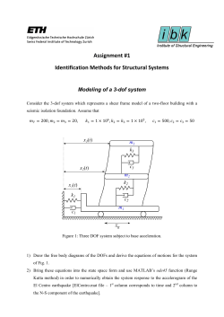
© Copyright 2026 ExpyDoc