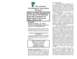
Epilepsy Research (2013) 107, 1—8
journal homepage: www.elsevier.com/locate/epilepsyres
REVIEW
Metabolic and endocrine effects of valproic
acid chronic treatment
DR
Vincenzo Belcastro a,∗, Claudia D’Egidio b, Pasquale Striano c,
Alberto Verrotti d
a
or
C
Neurology Unit, Department of Neuroscience, Sant’Anna Hospital, Como, Italy
Department of Pediatrics, University of Chieti, Chieti, Italy
c
Pediatric Neurology and Muscular Diseases Unit, Department of Neurosciences, Rehabilitation,
Ophtalmology, Genetics, Maternal and Child Health, University of Genoa, ‘‘G. Gaslini’’ Institute, Genova,
Italy
d
Department of Pediatrics, University of Perugia, Perugia, Italy
ap
b
ut
Co
pi
Valproic acid;
Epileptic syndromes;
Metabolic syndrome;
Atherosclerosis
Summary Treatment of epileptic patients with valproic acid (VPA) may be associated with
substantial weight changes that may increase morbidity and impair adherence to the treatment
regimen. VPA-induced weight gain seems to be associated with many metabolic disturbances;
the most frequent are hyperinsulinemia and insulin resistance, hyperleptinemia and leptin resistance. Patients who gain weight during VPA therapy can develop dyslipidemia and metabolic
syndrome that are associated with long-term vascular complications such as hypertension and
atherosclerosis. Moreover, an elevation in the levels of uric acid and homocysteine, together
with oxidative stress, may contribute to atherosclerotic risk in patients under long-term therapy
with VPA.
The aim of this review is to discuss the metabolic and endocrine effects of VPA chronic
treatment in patients with epilepsy.
© 2013 Elsevier B.V. All rights reserved.
aa
KEYWORDS
or
iz
ad
Received 19 February 2013 ; received in revised form 23 July 2013; accepted 14 August 2013
Available online 4 September 2013
Contents
Introduction ................................................................................................................
Valproic acid and weight gain...............................................................................................
Valproic acid and hyperinsulinaemia ........................................................................................
∗
Corresponding author. Tel.: +39 0 31 5859682; fax: +39 0 31 5859684.
E-mail addresses: [email protected], [email protected] (V. Belcastro).
0920-1211/$ — see front matter © 2013 Elsevier B.V. All rights reserved.
http://dx.doi.org/10.1016/j.eplepsyres.2013.08.016
15/09/2014
2
2
3
2
V. Belcastro et al.
Valproic acid and metabolic syndrome......................................................................................
Valproic acid and atherogenesis ............................................................................................
Conclusions.................................................................................................................
References .................................................................................................................
4
5
6
6
Valproic acid and weight gain
Anticonvulsant properties of valproic acid (VPA), which is
structurally unrelated to other antiepileptic drugs, were discovered by chance. The drug was first synthesized in 1882 by
Burton (1882) as an analogue of valeric acid, naturally found
in valerian (Burton, 1882). VPA is a fatty acid that is a clear
liquid at room temperature and, for many decades, its only
use was in laboratories as a solvent for organic compounds.
In 1962, Pierre Eymard, a research student at the University
of Lyon, used VPA as a solvent to investigate the potential anticonvulsant drugs with low aqueous solubility against
pentylenetetrazol-induced convulsions (PTZ) in laboratory
rats. A strong anticonvulsant activity was observed in all
solutions where the compound was present, leading to investigate VPA as a potentially useful agent for epilepsy (Meunier
et al., 1963). Thus, VPA was approved as an antiepileptic
drug in 1967 in France and it is currently the antiepileptic
drug (AED) with the broadest spectrum across all types of
seizures and epileptic syndromes (Aldenkamp et al., 2006;
Striano and Belcastro, 2012, 2013). Further, VPA was the
first, and, until the 1990, the only drug with a very broad
spectrum of activity and its efficacy has been seen in idiopathic generalized epilepsy with or without photosensitivity,
idiopathic focal and symptomatic generalized tonic-clonic
seizures (Aldenkamp et al., 2006). In addition, VPA has
gained acceptance in the treatment of bipolar disorder and
impulsive—aggressive behaviour in patients with personality
disorders other than in migraine prophylaxis (Johannessen
Landmark, 2008).
While the side effects of VPA are well documented in
clinical practice, little is known about the effects of chronic
VPA treatment. Given the heterogeneous prescribing patterns and varied benefits of VPA, the clarification of their
safety profile merits attention. VPA has been associated with
metabolic and endocrine disorders as weight gain and hyperinsulinaemia that may contribute to cardiovascular risk in
patients with epilepsy (Verrotti et al., 2010). Moreover, possibly influencing atherothrombotic risk factors such as serum
lipids, lipoprotein (a), uric acid level and homocysteine, VPA
treatment may represents an underling risk factor for systemic vascular diseases.
Despite its long-standing usage, the mechanism (s) of
the anticonvulsant activity of VPA is still controversial.
In fact, mechanistic studies originally focused on its ability to dampen neuronal hyperexcitability by potentiation
of inhibitory neurotransmission through an effect on ␥aminobutyric acid (GABA) metabolism, while more recent
studies have shown new activities for VPA including effects
on voltage-gated sodium channels, NMDA receptor-mediated
actions and as Histone deacetylase (HDAC) inhibitor.
This article reviews on the effects of VPA chronic treatment, focusing on metabolic and endocrine disorders and as
possible risk factor for atherosclerosis.
Significant weight gain has been one of the most frequently
experienced problems in patients with epilepsy (10—70%)
spanning paediatric to adult usage of VPA (Isojärvi et al.,
1996; Novak et al., 1999; Rättyä et al., 1999; Verrotti et al.,
1999, 2002, 2004, 2010; Pylvänen et al., 2002; El-Khayat
et al., 2004; de Vries et al., 2007; El-Khatib et al., 2007;
Hamed et al., 2009; Sharpe et al., 2009).
Among all these studies, weight change has not been
evaluated specifically in VPA clinical trials except in a
double-blind, 1 year study of new onset seizures comparing CBZ, topiramate, and VPA (Privitera et al., 2003). In this
study, patients receiving VPA increased their weight by an
average of 2.0 kg (2.8% of baseline weight) and 5.0 kg in children. Conversely, CBZ was weight neutral (Privitera et al.,
2003). In a double-blind study comparing lamotrigine (LTG)
and VPA, weight changes were assessed and after 32 weeks
of treatment, mean weight gain was significantly higher in
VPA-treated than LTG-treated patients (Biton et al., 2003).
With regard to the potential risk factors, results from
clinical studies have suggested that the occurrence of weight
gain is more prevalent in females with epilepsy than in males
(Sthephen et al., 2001; Hamed et al., 2009; Kanemura et al.,
2012), therefore, the gender may be considered one of risk
factors for VPA-induced weight gain. In particular, El-Khatib
et al. (2007) reported a significant weight gain in 43.6% of
women compared with 23.5% of men receiving VPA therapy.
Furthermore, percentage of body fat and waist-to-hip ratio
differed statistically between genders with women having
higher percentage of body fat and a lower waist/hip ratio.
From the analysis of these data, in female patients,
a further risk factor is the younger age: the increase
in body weight appears to occur more frequently in post
pubertal girls taking VPA (Rättyä et al., 1999; Verrotti et al.,
1999; Biton et al., 2003; Prabhakar et al., 2007; de Vries
et al., 2007) and body weight increase is more common in
patients treated with VPA during puberty if epilepsy and
therapy continue into adulthood (Mikkonen et al., 2005). In
adolescent girls, excessive weight gain has not only serious
psychological effects but can also causes the development
of important endocrinological abnormalities and a decrease
of treatment compliance. Then, this side effect must be
addressed if medication for females with epilepsy is begun
before 20 years of age. The mechanism through which VPA
may induce a weight gain is matter of discussion. However,
various hypotheses have been submitted to explain the
effect of VPA on weight increase: dysregulation of the
hypothalamic system, effect on adipokine levels, hyperinsulinaemia, IR. Experimental data have demonstrated that
VPA can cause dysregulation of the hypothalamic system
(Lakhanpal and Kau, 2007). This theory may be explained
by the enhancement of GABA transmission within the
hypothalamic axis (Biton et al., 2003) and it is supported
Co
pi
aa
ut
or
iz
ad
ap
or
C
DR
Introduction
15/09/2014
Metabolic and endocrine effects
3
children prior to and independent of a consistent weight
gain during the first years of VPA: so, a derangement in
ghrelin secretion in epilepsy during VPA treatment and
independent of weight gain could be hypothesized.
Valproic acid and hyperinsulinaemia
ap
or
C
DR
In general, hyperinsulinaemia is known to be associated with
obesity, dyslipidaemia and IR. Several studies in adults and
children have established that hyperinsulinemia occurs in
patients treated with VPA after the increase of body weight
(Rättyä et al., 1999; Verrotti et al., 1999; Sthephen et al.,
2001; Luef et al., 2002a,b; Pylvänen et al., 2002; Verrotti
et al., 2002; El-Khayat et al., 2004; Pylvänen et al., 2006;
Hamed et al., 2009; Kanemura et al., 2012).
Thus, it is likely that the VPA-induced weight gain is the
cause of hyperinsulinemia and IR (Verrotti et al., 2010) and,
consequently, patients who gain weight and develop IR after
VPA treatment may have a higher risk of metabolic MS than
patients without an increase of the body weight. On the
other hand, Isojärvi et al. firstly found that the development
of hyperinsulinemia and IR in long-term VPA therapy may
be among multiple factors leading to weight gain in some
patients. This observation is in agreement with data of successive studies that have suggested that hyperinsulinaemia
in obese patients taking VPA is not merely a consequence of
IR induced by weight gain but the development of IR may be
one of the factors leading to weight gain in some patients
(Pylvänen et al., 2002; Verrotti et al., 2002; Pylvänen et al.,
2003; Lihn et al., 2005; Pylvänen et al., 2006). This is
supported by the observation that weight gain during VPA
treatment is related to increase in insulin concurrent with
decrease in glucose level, which can stimulate appetite and
may cause weight gain (Demir and Aysun, 2000). Interestingly, Pylvänen et al. (2006) have tried to explain as VPA
may also determine hyperinsulinaemia in lean patients: they
studied 51 adult patients on VPA monotherapy and compared
them with 45 healthy control subjects with respect to fasting
plasma glucose, serum insulin, proinsulin and C-peptide concentrations after overnight fast. The VPA-treated patients
had fasting hyperinsulinaemia, although the fasting serum
proinsulin and C-peptide concentrations were not significantly higher compared with the control. Therefore, VPA
could not induce insulin secretion but may interfere with
insulin metabolism in the liver, resulting in higher insulin
concentrations in peripheral circulation. With regard to the
mechanisms by which VPA may induce IR, other hypotheses
have been proposed such as the increased plasma levels of
free fatty acids (FFA) caused by VPA (McGarry, 2002; Luef
et al., 2002a,b), b-cell dysfunction as a consequence of the
oxidative stress (Evans et al., 2003), a direct effect on bcells regulation and insulin secretion (Luef et al., 2009),
and finally, an alteration of the sympathetic nervous system by acting on hypothalamic neurons (Breum et al., 1992).
Recently, retinol-binding protein 4 (RBP4) and Glucagon-like
peptide-1 (GLP-1) are considered as important new targets
in modern type 2 diabetes mellitus therapy linked to IR and
nonalcoholic fatty liver disease (NAFLD); Rauchenzaunera
et al. (2012) have demonstrated the lack of an influence
of VPA treatment on RBP4 and GLP-1 in otherwise healthy
patients. In summary, the absence of any relationship with
Co
pi
aa
ut
or
iz
ad
by the observation that VPA-treated epileptic patients
who reported weight gain developed increased appetite
and quenching with calorie-rich beverages (Verrotti et al.,
1999). A more recent hypothesis is that VPA may induce
weight gain by the modifying expression of adipokine genes
that are expressed in the brain and pituitary (cephalokines);
these genes codify for neuropeptides involved in central
energy metabolism, such as resistin and fasting-induced adipose factor also known as angiopoietin-like protein 4, which
have become major targets implicated in the aetiology of
obesity and development of leptin and insulin resistance
(IR) (Brown et al., 2008). Although VPA may modify hypothalamic gene expression in vitro (Münzberg and Myers, 2005),
it is unclear whether it has similar effects in vivo.
VPA may have effects on adipokine released by adipose tissue, such as adiponectin, leptin, soluble leptin
receptor and on ghrelin. Ghrelin is the natural ligand of
the growth hormone secretagogue receptor. In fact, VPA
can suppress adiponectin gene expression in adipocytes
through HDAC inhibition (Qiao et al., 2006). These findings
are in agreement with data showing lower concentrations
of adiponectin in patients with obesity and type 2 Diabetes, providing a clear evidence of a relationship between
overweight and overweight-related disorders (Greco et al.,
2005). Interestingly, VPA can increase the expression of
mRNA of adiponectin-binding receptors, adipoR1, in human
hepatoma cell line HepG2 cells (Rauchenzauner et al.,
2008a). As adiponectin mRNA expression is known to be
downregulated following VPA treatment in vivo (Greco et al.,
2005) and in vitro (Qiao et al., 2006), increased adipoR1
mRNA expression in liver cells possibly represents a favoured
reaction balancing suppressed adiponectin secretion from
adipocytes; changes in this balance of receptor/ligand
expression might contribute to changes in fatty acid oxidation and IR in VPA-related obesity.
On the other side, hyperleptinaemia and leptin resistance
are associated with obesity; consequently, this condition
can explain the VPA-induced weight gain: clinical studies
reported increased serum levels of leptin in children and
adults who gain weight during VPA treatment (Aydin et al.,
2005, Hamed et al., 2009; Rauchenzauner et al., 2008b). The
effects of VPA on leptin biology and fatty acid metabolism
have been tested in 3T3-L1 adipocytes (Rauchenzauner
et al., 2008c): in vitro, VPA paradoxically reduces leptin
mRNA levels and secretion of the leptin protein in a doseand time-dependent manner. Probably, the inhibition of leptin secretion by VPA induces enhanced appetite in patients,
resulting in enhanced adiposity and an increase in leptin
secretion (see Fig. 1; Table 1).
Finally, because an association between ghrelin levels
and obesity has been shown in humans (ghrelin levels
are reduced in obesity), several studies have studied the
relationship between VPA treatment and ghrelin: it seems
that ghrelin levels are reduced in VPA-induced obesity (van
der Lely et al., 2004; Greco et al., 2005; Ness-Abramof
and Apovian, 2005; Gungor et al., 2007; Prodam et al.,
2012). It is known that ghrelin influences glucose and insulin
metabolism and the control of food and energy intake
through the neuropeptide Y (NPY) system. Furthermore,
ghrelin levels are reduced in obesity (van der Lely et al.,
2004). Interestingly, Prodam et al. (2012) showed that
ghrelin levels are decreased in very young prepubertal
15/09/2014
4
V. Belcastro et al.
Pathogenetic mechanisms of VPA-induced weight.
prospective study that focused on VPA-treated children and
adolescents; our study showed that 40.4% of the patients
exhibited considerably body weight, whereas 43.5% of the
obese patients were diagnosed with MS at the end of the 24
months of follow-up (Verrotti et al., 2010). A study evaluating the presence of MS among Chinese adult obese patients
with epilepsy on VPA therapy suggests that obese patients
with epilepsy treated with VPA are at higher risk of MS than
individuals who are simply obese (Fang et al., 2012); this
study also demonstrated the homeostatic model assessment
(HOMA) index is related to MS development rather than
body-mass index. Thus, the HOMA index should be monitored
in obese VPA-treated patients routinely (Fang et al., 2012).
In addition, the possible relevant factors for MS development among patients were determined: MS was associated
with high VPA doses but was independent of age, gender,
seizure type, and duration of medication. This finding has to
be considered an important matter, as the necessary dosage
might be adjusted to minimize the possibility of occurrence
of MS attributed to VPA treatment.
In the last years, it has been demonstrated that IR may
be related to the development of non-alcoholic fatty liver
disease (NAFLD) (Angulo, 2002). NAFLD can be considered a
ap
or
C
RBP4 and GLP-1 concentrations does not suggest a role of
these novel IR parameters as potential regulators of glucose
and fat metabolism during VPA-therapy.
VPA: valproic acid; ALP-4: angiopoietin-like protein.
DR
Fig. 1
ad
Valproic acid and metabolic syndrome
Co
pi
aa
ut
or
iz
Metabolic syndrome (MS) is a constellation of metabolic risk
factors that includes increased waist circumference, atherogenic dyslipidemia, elevated blood pressure, and elevated
blood glucose associated with IR (Alberti et al., 2006). Several meta-analyses have shown that MS is associated with
an approximately 2-fold increased risk of cardiovascular disease (Isomaa et al., 2001; Lakka et al., 2002; Mottillo et al.,
2010).
Recent studies have revealed that MS represents an enormous economic burden and is considered a serious public
health problem (Fu et al., 2007; Wang et al., 2010). High
prevalence of MS has been reported among patients with
bipolar disorder who experienced significant weight gain
associated with VPA (de Almeida et al., 2012). However,
the presence of MS among obese patients with epilepsy
on VPA has received little attention. We have conducted a
Table 1
Summary of the potential metabolic and endocrine effects of valproic acid chronic treatment.
Effect
Risk factor
Mechanism
Weight gain
Female, young age
Hyperinsulinaemia
VPA-induced weight gain
Insulin resistance
VPA-induced weight gain
Metabolic syndrome
Atherogenesis
High VPA doses
Dyslipidemia, hyper-Hcy high
uric acid level gain.
Dysregulation of the hypothalamic system,
effect on adipokine levels, hyperinsulinaemia
VPA interferes with insulin metabolism in the
liver
Increased plasma levels of FFA, b-cell
dysfunction increased insulin secretion
IR, NAFLD
Dysfunction of the vessel wall
IR: insulin resistance; VPA: valproic acid; FFA: free fatty acids; NAFLD: non-alcoholic fatty liver disease; Hcy: homocysteine.
15/09/2014
Metabolic and endocrine effects
5
Co
pi
aa
ut
DR
or
iz
ad
Atherosclerosis is the leading cause of death in the developed world, although its true frequency is difficult to be
accurately determined because it is a predominantly asymptomatic condition (Berenson et al., 1998). Interestingly,
epidemiological studies have indicated that the prevalence
and death rates from atherosclerosis related cardiovascular
disease are elevated in adult epileptic patients (Annegers
et al., 1984; Gaitatzis et al., 2004). Influence of AEDs on
the development of atherosclerosis has been the subject
of controversy; in fact, recent evidence indicates that prolonged antiepileptic treatment might modify some vascular
risk factors (Hamed and Nabeshima, 2005; Hamed et al.,
2007; Elliott et al., 2007) while other studies showed that
the mortality due to ischaemic heart disease appears to be
lower in treated epileptics than in the general population
(Kaste et al., 1983; Muuronen et al., 1985; Olesen et al.,
2011). Recent studies have provided evidence that chronic
administration of older AEDs is associated with the undesirable metabolic side effects (Mintzer and Mattson, 2009;
Brodie et al., 2013) implicated in dysfunction of the vessel
wall (Tan et al., 2009), the key pathophysiological mechanism promoting atherosclerosis.
It is well established that an increased carotid artery
intima media thickness (CA-IMT) is a good predictor of clinical manifestation of atherosclerosis (Polak et al., 2011).
Noteworthy, several studies of carotid arteries in patients
with epilepsy have demonstrated significantly increased CAIMT relative to normal controls (Hamed et al., 2007; Tan
et al., 2009; Chuang et al., 2012). Interestingly, Erdemir
et al. (2009) found an increased CA-IMT in epileptic children
treated with VPA, while in a similar manner, the authors
failed to found an increased CA-IMT in epileptic children
treated with oxcarbazepine (Yis¸ and Do˘
gan, 2012).
The first signs of hyperlipidemia can be detected in childhood (Strong et al., 2001), and fatty streaks, which are
the earliest pathologic lesions of the atherogenic process,
can be observed in the arteries of individuals by the age
of 20 years (Berenson et al., 1998). Dyslipidemia has long
been known to be an important risk factor for atherosclerosis (Kullo and Ballantyne, 2005). Low density lipoproteins
(LDLs) plays an important role in the atherosclerotic process by increasing endothelial permeability, retention of
lipoproteins within the intima of blood vessels, recruitment of inflammatory cells and formation of foam cells
(Stocker and Keaney, 2004; Kullo and Ballantyne, 2005).
Emerging evidence showed that treatment with enzymeinducing AEDs, such as carbamazepine (CBZ) and phenytoin
(PHT) is significantly associated with increased blood levels
of total cholesterol, atherogenic (non-HDL) cholesterol and
or
C
Valproic acid and atherogenesis
triglycerides. Probably, the increase in thickness of CCA IMT
in patients treated with PHT or CBZ may be related to total
cholesterol and LDLs (Chuang et al., 2012; Sonmez et al.,
2006; Tomoum et al., 2008). Instead, the effects of VPA
on changes in lipid profiles and lipoproteins remains controversial (Eirìs et al., 1995; Geda et al., 2002; Nikolaos
et al., 2004; Pylvänen et al., 2006; Abaci et al., 2009;
Grosso et al., 2009; Lopinto-Khoury and Mintzer, 2010;
Chuang et al., 2012): some studies (Geda et al., 2002;
Nikolaos et al., 2004; Chuang et al., 2012) found no effect
on plasma concentrations of total cholesterol, high-density
lipoprotein cholesterol, or its components, whereas others
demonstrated significant changes in lipids, lipoproteins, and
apolipoproteins (Demircioglu et al., 2000; Voudris et al.,
2006; Abaci et al., 2009; Grosso et al., 2009; Verrotti et al.,
2010); in particular, Abaci et al. found a significant increase
in total cholesterol and LDLs after 12 months of VPA treatment, but triglycerides and HDLs levels did not change.
High serum triglyceride concentrations and low HDL were
found in patients on VPA treatment which have developed
the MS, and there were no significant differences by gender (Verrotti et al., 2010). In this work, the dyslipidemia
was associated with the IR; in fact, it is known that hyperinsulinemia increases lipogenesis that can be responsible for
the accumulation of triglycerides: insulin plays a central
role in determining triglyceride clearance from the blood
via activation of lipoprotein lipase and triglyceride output through effects on the synthesis and secretion of very
LDL (VLDL) by the liver (Lewis and Steiner, 1996). Furthermore, insulin controls the output of free fatty acids from
adipose tissue (Arner, 1995); consequently, a state of IR
may determine a delay in plasma lipoprotein triglyceride
clearance, that allows for cholesterol esters to be passed
on from HDL to triglyceride-rich particles, which results in
potentially atherogenic lipoprotein particles (Patsch et al.,
1992).
Homocysteine (Hcy) is a sulfur-containing, nonprotein amino acid reversibly formed and secreted during
metabolism of methionine. Once formed, Hcy is metabolized via two pathways: (i) re-methylation to methionine, which requires methylenetetrahydrofolate reductase
(MTHFR)/methionine synthase (MS) or betaine homocysteine methyltransferase (BHMT), and folic acid and vitamin
B12 as co-factors; (ii) trans-sulfuration to cysteine, which
requires cystathionine-beta-synthase (CBS) and pyridoxal5 -phosphate, the vitamin B6 coenzyme (Mattson and Shea,
2003; Belcastro et al., 2007; Belcastro et al., 2010). Epidemiological data demonstrated that elevated hyper-Hcy
concentration is an independent risk factor for the progression of atherosclerosis (Temple et al., 2000; Hassan et al.,
2004; Belcastro and Striano, 2012). In particular, hyper-Hcy
has been associated with cardiovascular disease and stroke
in multiple large-scale epidemiologic studies (Belcastro and
Striano, 2012). Notably, it has been showed that, among
older AEDs, prolonged treatment with VPA raises tHcy levels
in epileptic children (Verrotti et al., 2000; Karabiber et al.,
2003; Attilakos et al., 2006).
Finally, it has been shown that uric acid level was significantly higher in patients with VPA monotherapy; it is known
that higher levels of serum uric acid may be associated with
the development of atherosclerosis that is independent of
other atherosclerotic risk factors.
ap
feature of the metabolic syndrome (Marchesini et al., 2001),
associated with the presence of IR. In three clinical studies
(Stiemer, 1989; Luef et al., 2004, 2009), ultrasound examination of the liver in patients affected by epilepsy revealed
that 61% of patients on VPA and 21% with CBZ had NAFLD.
However, taking into account that NAFLD may be dangerous because it can be considered a component of the MS
and it can be followed by cirrhosis, an early diagnosis is
essential.
15/09/2014
6
V. Belcastro et al.
pi
References
aa
ut
or
C
ap
or
iz
ad
Since its first marketing as an antiepileptic drug more
than 45 years ago in France, VPA has become established
worldwide as one of the most widely used AEDs in the
treatment of both generalized and partial seizures in adults
and children. The broad spectrum of antiepileptic efficacy of VPA is reflected in preclinical in vivo and in vitro
models, including a variety of animal models of seizures
or epilepsy. VPA may have many adverse effects (usually
considered idiosyncratic) that require vigilance during the
chronic treatment: in particular, its use is not recommended
in patients with some preexisting conditions e.g. hepatic and
pancreatic insufficiency; moreover, VPA is clearly associated
with weight changes and related endocrine abnormalities:
therefore, this drug should be avoided in obese patients
(at risk of developing MS), in particular in female pubertal
patients because VPA can be expected to cause polycystic
ovaries. Moreover, increase weight may account for increase
of atherosclerosis risks: consequently, this drug should be
avoided in a familiar context of atherosclerosis and/or cardiovascular diseases.
There is no single mechanism of action of this AED that
can account for all the numerous effects of the drug on
neuronal tissue and its broad clinical activity. Furthermore,
by the experimental and clinical observations summarized
in this review, the potential many endocrinological and
metabolic side effects of VPA remain to be explained.
Changes involving the levels of key molecules or neurotransmitters such as insulin, leptin, neuropeptide, ghrelin, and
adiponectin may influence weight gain and VPA-induced obesity with the subsequent metabolic consequences. In view of
the advances in molecular neurobiology and neuroscience,
future studies will hopefully further our understanding of
the mechanisms of action of this amazing drug.
Belcastro, V., Gorgone, G., Italiano, D., Oteri, G., Caccamo,
D., Pisani, L.R., et al., 2007. Antiepileptic drugs and MTHFR
polymorphisms influence hyper-homocysteinemia recurrence in
epileptic patients. Epilepsia 48, 1990—1994.
Belcastro, V., Striano, P., Gorgone, G., Costa, C., Ciampa, C.,
Caccamo, D., et al., 2010. Hyperhomocysteinemia in epileptic
patients on new antiepileptic drugs. Epilepsia 51, 274—279.
Belcastro, V., Striano, P., 2012. Antiepileptic drugs, hyperhomocysteinemia and B-vitamins supplementation in patients with
epilepsy. Epilepsy Res. 102, 1—7.
Berenson, G.S., Srinivasan, S.R., Bao, W., Newman, W.P., Tracy,
R.E., Wattigney, W.A., 1998. Association between multiple cardiovascular risk factors and atherosclerosis in children and young
adults. The Bogalusa Heart Study. N Engl J Med. 338, 1650—1656.
Biton, V., Levisohn, P., Hoyler, S., Vuong, A., Hammer, A.E., 2003.
Lamotrigine versus valproate monotherapy-associated weight
change in adolescents with epilepsy: results from a post hoc
analysis of a randomized, double-blind clinical trial. J. Child
Neurol. 18, 133—139.
Breum, L., Astrup, A., Gram, L., Andersen, T., Stokholm, K.H.,
Christensen, N.J., et al., 1992. Metabolic changes during treatment with valproate in humans: implication for untoward weight
gain. Metab. Clin. Exp. 41, 666—670.
Brodie, M.J., Mintzer, S., Pack, A.M., Gidal, B.E., Vecht, C.J.,
Schmidt, D., 2013. Enzyme induction with antiepileptic drugs:
cause for concern? Epilepsia 54, 11—27.
Brown, R., Imran, S.A., Ur, E., Wilkinson, M., 2008. Valproic acid and
CEBPalpha-mediated regulation of adipokine gene expression in
hypothalamic neurons and 3T3-L1 adipocytes. Neuroendocrinology 88, 25—34.
Burton, B.S., 1882. On the propyl derivatives and decomposition
products of ethylacetoacetate. Am. Chem. J. 3, 385—395.
Chuang, Y.C., Chuang, H.Y., Lin, T.K., Chang, C.C., Lu, C.H.,
Chang, W.N., et al., 2012. Effects of long-term antiepileptic
drug monotherapy on vascular risk factors and atherosclerosis.
Epilepsia 53, 120—128.
de Almeida, K.M., Moreira, C.L., Lafer, B., 2012. Metabolic syndrome and bipolar disorder: what should psychiatrists know? CNS
Neurosci. Ther. 18, 160—166.
de Vries, L., Karasik, A., Landau, Z., Phillip, M., Kiviti, S., GoldbergStern, H., 2007. Endocrine effects of valproate in adolescent
girls with epilepsy. Epilepsia 48, 470—477.
Demir, E., Aysun, S., 2000. Weight gain associated with valproate in
childhood. Pediatr. Neurol. 22, 361—364.
Demircioglu, S., Soylu, A., Dirik, E., 2000. Carbamazepine and valproic acid: effects on the serum lipids and liver functions in
children. Pediatr. Neurol. 23, 142—146.
Eirìs, J.M., Lojo, S., Del Rìo, M.C., Novo, I., Bravo, M., Pavon, P.,
et al., 1995. Effects of long-term treatment with antiepileptic
drugs on serum lipid levels in children with epilepsy. Neurology
45, 1155—1157.
Elliott, J.O., Jacobson, M.P., Haneef, Z., 2007. Cardiovascular risk
factors and homocysteine in epilepsy. Epilepsy Res. 76, 113—123.
El-Khayat, H.A., El-Basset, F.Z., Tomoum, H.Y., Tohamy, S.M.,
Zaky, A.A., Mohamed, M.S., et al., 2004. Physical growth and
endocrinal disorders during pubertal maturation in girls with
epilepsy. Epilepsia 45, 1106—1115.
El-Khatib, F., Rauchenzauner, M., Lechleitner, M., Hoppichler, F.,
Naser, A., Waldmann, M., et al., 2007. Valproate, weight
gain and carbohydrate craving: a gender study. Seizure 16,
226—232.
glu, F., Kir, M., Cakmakc
¸i, H.,
Erdemir, A., Cullu, N., Yis, U., Demircio˘
et al., 2009. Evaluation of serum lipids and carotid artery intima
media thickness in epileptic children treated with valproic acid.
Brain Dev. 31, 713—716.
Evans, J.L., Goldfine, I.D., Maddux, B.A., Grodsky, G.M., 2003.
Are oxidative stress-activated signaling pathways mediators of
insulin resistance and beta-cell dysfunction? Diabetes 52, 1—8.
DR
Conclusions
Co
Abaci, A., Saygi, M., Yis, U., Demir, K., Dirik, E., Bober, E., 2009.
Metabolic alterations during valproic acid treatment: a prospective study. Pediatr. Neurol. 41, 435—439.
Alberti, K.G., Zimmet, P., Shaw, J., 2006. Metabolic syndrome —
a new world-wide definition. A consensus statement from the
International Diabetes Federation. Diabet. Med. 23, 469—480.
Angulo, P., 2002. Nonalcoholic fatty liver disease. N. Engl. J. Med.
346, 1221—1231.
Aldenkamp, A., Vigevano, F., Arzimanoglou, A., Covanis, A., 2006.
Role of valproate across the ages. Treatment of epilepsy in children. Acta Neurol. Scand. 184, 1—13.
Annegers, J.F., Hauser, W.A., Shirts, S.B., 1984. Heart disease mortality and morbidity in patients with epilepsy. Epilepsia 25,
699—704.
Arner, P., 1995. Differences in lipolysis between human subcutaneous and omental adipose tissue. Ann. Med. 27, 435—438.
Attilakos, A., Papakonstantinou, E., Schulpis, K., Voudris, K., Katsarou, E., Mastroyianni, et al., 2006. Early effect of sodium
valproate and carbamazepine monotherapy on homocysteine
metabolism in children with epilepsy. Epilepsy Res. 71, 229—232.
Aydin, K., Serdaroglu, A., Okuyaz, C., Bideci, A., Gucuyener,
K., 2005. Serum insulin, leptin, and neuropeptide y levels in
epileptic children treated with valproate. J. Child Neurol. 20,
848—851.
15/09/2014
Metabolic and endocrine effects
7
ap
or
C
DR
Lewis, G.F., Steiner, G., 1996. Acute effects of insulin in the control
of VLDL production in humans. Implications for the insulinresistant state. Diabetes Care 19, 390—393.
Lihn, A.S., Pedersen, S.B., Richelsen, B., 2005. Adiponectin: action,
regulation and association to insulin sensitivity. Obes. Rev. 6,
13—21.
Lopinto-Khoury, C., Mintzer, S., 2010. Antiepileptic drugs and markers of vascular risk. Curr. Treat. Options Neurol. 12, 300—308.
Luef, G., Abraham, I., Trinka, E., Alge, A., Windisch, J., Daxenbichler, G., et al., 2002a. Hyperandrogenism, postprandial
hyperinsulinism and the risk of PCOS in a cross-sectional study
of women with epilepsy treated with valproate. Epilepsy Res. 2
(48), 91—102.
Luef, G., Abraham, I., Hoppichler, F., Trinka, E., Unterberger, I.,
Bauer, G., et al., 2002b. Increase in postprandial serum insulin
levels in epileptic patients with valproic acid therapy. Metab.
Clin. Exp. 51, 1274—1278.
Luef, G., Waldmann, M., Sturm, W., Naser, A., Trinka, E., Unterberger, I., et al., 2004. Valproate therapy and non-alcoholic fatty
liver disease. Ann. Neurol. 55, 729—732.
Luef, G., Rauchenzauner, M., Waldmann, M., Sturm, W., Sandhofer,
A., Seppi, K., et al., 2009. Non-alcoholic fatty liver disease
(NAFLD), insulin resistance and lipid profile in antiepileptic drug
treatment. Epilepsy Res. 86, 42—47.
Marchesini, G., Brizi, M., Bianchi, G., Tomassetti, S., Bugianesi, E.,
Lenzi, M., et al., 2001. Nonalcoholic fatty liver disease: a feature
of the metabolic syndrome. Diabetes 50, 1844—1850.
Mattson, M.P., Shea, T.B., 2003. Folate and homocysteine
metabolism in neural plasticity and neurodegenerative disorders. Trends Neurosci. 26, 137—146.
McGarry, J.D., 2002. Dysregulation of fatty acid metabolism in the
etiology of type 2 diabetes. Diabetes 51, 7—18.
Meunier, H., Carraz, G., Neunier, Y., Eymard, P., Aimard, M., 1963.
Pharmacodynamic properties of ndipropylacetic acid. Therapie
18, 435—438.
Mikkonen, K., Knip, M., Pakarinen, A.J., Lanning, P., Isojärvi, J.,
Vainionpää, L.K., 2005. Growth and lipid metabolism in girls and
young women with epilepsy during pubertal maturation. Epilepsia 46, 1114—1120.
Mintzer, S., Mattson, R.T., 2009. Should enzyme-inducing antiepileptic drugs be considered first-line agents? Epilepsia 50, 42—50.
Mottillo, S., Filion, K.B., Genest, J., Joseph, L., Pilote, L., Poirier,
P., et al., 2010. The metabolic syndrome and cardiovascular risk
a systematic review and meta-analysis. J. Am. Coll. Cardiol. 56,
1113—1132.
Münzberg, H., Myers, M.G., 2005. Molecular and anatomical determinants of central leptin resistance. Nat. Neurosci. 8, 566—570.
Muuronen, A., Kaste, M., Nikkila, E.A., Tolppanen, E.M., 1985. Mortality from ischemic heart disease among the patients using
anticonvulsant drugs. A case control study. BMJ 291, 1481—1483.
Ness-Abramof, R., Apovian, C.M., 2005. Drug-induced weight gain.
Drugs Today 41, 547—555.
Nikolaos, T., Stylianos, G., Chryssoula, N., Irini, P., christos, M.,
Dimitrios, T., 2004. The effect of long-term antiepileptic treatment on serum cholesterol (TC, HDL, LDL) and triglycerid elevels
in adult epileptic patients on monotherapy. Med. Sci. Monit. 10,
50—52.
Novak, G.P., Maytal, J., Alshansky, A., Eviatar, L., Sy-Kho, R.,
Siddique, Q., 1999. Risk of excessive weight gain in epileptic
children treated with valproate. J. Child Neurol. 14, 490—495.
Olesen, J.B., Hansen, P.R., Abildstrøm, S.Z., Andersson, C., Weeke,
P., Schmiegelow, M., et al., 2011. Valproate attenuates the risk
of myocardial infarction in patients with epilepsy: a nationwide
cohort study. Pharmacoepidemiol. Drug. Saf. 20, 146—153.
Patsch, J.R., Miesenbock, G., Hopferwieser, T., Muhlberger, V.,
Knapp, E., Dunn, J.K., et al., 1992. Relation of triglyceride
metabolism and coronary artery disease. Studies in the postprandial state. Arterioscler. Thromb. 12, 136—141.
Co
pi
aa
ut
or
iz
ad
Fang, J., Chen, S., Tong, N., Chen, L., An, D., Mu, J., et al., 2012.
Metabolic syndrome among Chinese obese patients with epilepsy
on sodium valproate. Seizure 21, 578—582.
Fu, J.F., Liang, L., Zou, C.C., Hong, F., Wang, C.L., Wang, X.M.,
et al., 2007. Prevalence of the metabolic syndrome in Zhejiang
Chinese obese children and adolescents and the effect of metformin combined with lifestyle intervention. Int. J. Obes. 31,
15—22.
Geda, G., Caksen, H., Icagasioglu, D., 2002. Serum lipids, vitamin B12 and folic acid levels in children receiving long-term
valproate therapy. Acta Neurol. Belg. 102, 122—126.
Greco, R., Latini, G., Chiarelli, F., Iannetti, P., Verrotti, A., 2005.
Leptin, ghrelin, and adiponectin in epileptic patients treated
with valproic acid. Neurology 65, 1808—1809.
Grosso, S., Mostardini, R., Piccini, B., Balestri, P., 2009. Body
mass index and serum lipid changes during treatment with valproic acid in children with epilepsy. Ann. Pharmacother. 43,
45—50.
Gungor, S., Yücel, G., Akinci, A., Tabel, Y., Ozerol, I.H., Yologlu, S.,
2007. The role of ghrelin in weight gain and growth in epileptic
children using valproate. J. Child Neurol. 22, 1384—1388.
Hamed, S.A., Nabeshima, T., 2005. The high atherosclerotic risk
among epileptics: the atheroprotective role of multivitamins. J.
Pharmacol. Sci. 98, 340—353.
Hamed, S.A., Hamed, E.A., Hamdy, R., Nabeshima, T., 2007. Vascular risk factors and oxidative stress as independent predictors
of asymptomatic atherosclerosis in adult patients with epilepsy.
Epilepsy Res. 74, 183—192.
Hamed, S.A., Fida, N.M., Hamed, E.A., 2009. States of serum leptin
and insulin in children with epilepsy: risk predictors of weight
gain. Eur. J. Paediatr. Neurol. 13, 261—268.
Hassan, A., Hunt, B.J., O’Sullivan, M., Bell, R., D’Souza, R., Jeffery, S., et al., 2004. Homocysteine is a risk factor for cerebral
small vessel disease, acting via endothelial dysfunction. Brain
127, 212—219.
Gaitatzis, A., Carroll, K., Majeed, A.W., Sander, J., 2004. The
epidemiology of the comorbidity of epilepsy in the general population. Epilepsia 45, 1613—1622.
Isojärvi, J.I., Laatikainen, T.J., Knip, M., Pakarinen, A.J., Juntunen,
K.T., Myllylä, V.V., 1996. Obesity and endocrine disorders in
women taking valproate for epilepsy. Ann. Neurol. 39, 579—584.
Isomaa, B., Almgren, P., Tuomi, T., Forsen, B., Lahti, K., Nissen, M.,
et al., 2001. Cardiovascular morbidity and mortality associated
with the metabolic syndrome. Diabetes Care 24, 683—689.
Johannessen Landmark, C., 2008. Antiepileptic drugs in nonepilepsy disorders: relations between mechanisms of action and
clinical efficacy. CNS Drugs 22, 27—47.
Kanemura, H., Sano, F., Maeda, Y., Sugita, K., Aihara, M., 2012.
Valproate sodium enhances body weight gain in patients with
childhood epilepsy: a pathogenic mechanisms and open-label
clinical trial of behavior therapy. Seizure 21, 496—500.
Karabiber, H., Sonmezgoz, E., Ozerol, E., Yakinci, C., Otlu, B.,
Yologlu, S., 2003. Effects of valproate and carbamazepine on
serum levels of homocysteine, vitamin B12, and folic acid. Brain
Dev. 25, 113—115.
Kaste, M., Muuronen, A., Nikkilä, E.A., Neuvonen, P.J., 1983.
Increase of low serum concentrations of high-density lipoprotein (HDL) cholesterol in TIA-patients treated with phenytoin.
Stroke 14, 525—530.
Kullo, I.J., Ballantyne, C.M., 2005. Conditional risk factors for
atherosclerosis. Mayo Clin. Proc. 80, 219—230.
Lakhanpal, D., Kau, G., 2007. Valproic acid alters GnRH-GABA
interactions in cycling female rats. Cell. Mol. Neurobiol. 27,
1069—1083.
Lakka, H.M., Laaksonen, D.E., Lakka, T.A., Niskanen, L.K., Kumpusalo, E., Tuomilehto, J., et al., 2002. The metabolic syndrome
and total and cardiovascular disease mortality in middle-aged
men. JAMA 288, 2709—2716.
15/09/2014
8
V. Belcastro et al.
ap
or
C
DR
with sodium valproate or lamotrigine monotherapy. Epilepsia 42,
1002—1006.
Stiemer, B., 1989. Morphological evaluation of steatosis in monolayer cultures (MDCK cells) after treatment with gentamicin and
valproic acid. Histol. Histopathol. 4, 421—428.
Stocker, R., Keaney, J.F., 2004. Role of oxidative modifications in
atherosclerosis. Physiol. Rev. 84, 1381—1478.
Strong, J.P., Zieske, A.W., Malcom, G.T., 2001. Lipoproteins and
atherosclerosis in children: an early marriage? Nutr. Metab. Cardiovasc. Dis. 11, 16—22.
Striano, P., Belcastro, V., 2012. Treatment of myoclonic seizures.
Expert Rev. Neurother. 12, 1411—1418.
Striano, P., Belcastro, V., 2013. Treating myoclonic epilepsy in
children: state of the art. Expert Opin. Pharmacother. 14,
1355—1361.
Tan, T.Y., Lu, C.H., Chuang, H.Y., Lin, T.K., Liou, C.W., Chang, W.N.,
et al., 2009. Long-term antiepileptic drug therapy contributes to
the acceleration of atherosclerosis. Epilepsia 50, 1579—1586.
Temple, M.E., Luzier, A.B., Kazierad, D.J., 2000. Homocysteine as
a risk factor for atherosclerosis. Ann. Pharmacother. 34, 57—65.
Tomoum, H.Y., Awadallah, M.M., Fouad, D.A., Ali, A.H., 2008. Lipid
profile, apolipoproteins A and B in children with epilepsy. J. Child
Neurol. 23, 1275—1281.
van der Lely, A.J., Tschöp, M., Heiman, M.L., Ghigo, E., 2004. Biological, physiological, pathophysiological, and pharmacological
aspects of ghrelin. Endocr. Rev. 25, 426—457.
Verrotti, A., Basciani, F., Morresi, S., de Martino, M., Morgese, G.,
Chiarelli, F., 1999. Serum leptin changes in epileptic patients
who gain weight after therapy with valproic acid. Neurology 53,
230—232.
Verrotti, A., Pascarella, R., Trotta, D., Giuva, T., Morgese, G.,
Chiarelli, F., 2000. Hyperhomocysteinemia in children treated
with sodium valproate and carbamazepine. Epilepsy Res. 41,
253—257.
Verrotti, A., Basciani, F., De Simone, M., Trotta, D., Morgese, G.,
Chiarelli, F., 2002. Insulin resistance in epileptic girls who gain
weight after therapy with valproic acid. J. Child Neurol. 17,
265—268.
Verrotti, A., Greco, R., Latini, G., De Simone, M., Chiarelli, F.,
2004. Obesity and plasma concentrations of alpha-tocopherol
and beta-carotene in epileptic girls treated with valproate. Neuroendocrinology 79, 157—162.
Verrotti, A., Manco, R., Agostinelli, S., Coppola, G., Chiarelli, F.,
2010. The metabolic syndrome in overweight epileptic patients
treated with valproic acid. Epilepsia 51, 268—273.
Voudris, K.A., Attilakos, A., Katsarou, E., et al., 2006. Early and
persistent increase in serum lipoprotein (a) concentrations in
epileptic children treated with carbamazepine and sodium valproate monother-apy. Epilepsy Res. 70, 211—217.
Wang, B., Liu, Y., He, P., Dong, B., Ou Yang, L., Ma, Y., et al., 2010.
Prevalence of metabolic syndrome in an elderly Chinese population: a community-based cross-sectional study. J. Am. Geriatr.
Soc. 58, 2027—2028.
Yis¸, U., Do˘
gan, M., 2012. Effects of oxcarbazepine treatment on
serum lipids and carotid intima media thickness in children.
Brain Dev. 34, 185—188.
Co
pi
aa
ut
or
iz
ad
Polak, J.F., Pencina, M.J., Pencina, K.M., O’Donnell, C.J., Wolf,
P.A., D’Agostino, R.B., 2011. Carotid-wall intima-media thickness and cardiovascular events. N. Engl. J. Med. 365, 213—221.
Privitera, M.D., Brodie, M.J., Mattson, R.H., Chadwick, D.W., Neto,
W., Wang, S., et al., 2003. Topiramate, carbamazepine and
valproate monotherapy: double-blind comparison in newly diagnosed epilepsy. Acta Neurol. Scand. 107, 165—175.
Prabhakar, S., Sahota, P., Kharbanda, P.S., Siali, R., Jain, V., Lal, V.,
2007. Sodium valproate, hyperandrogenism and altered ovarian
function in Indian women with epilepsy: a prospective study.
Epilepsia 48, 1371—1377.
Prodam, F., Ricotti, R., Genoni, G., Parlamento, S., Petri, A.,
Balossini, C., et al., 2012. Comparison of two classifications of
metabolic syndrome in the pediatric population and the impact
of cholesterol. J. Endocrinol. Invest..
Pylvänen, V., Knip, M., Pakarinen, A., Kotila, M., Turkka, J.,
Isojärvi, J.I., 2002. Serum insulin and leptin levels in valproateassociated obesity. Epilepsia 43, 514—517.
Pylvänen, V., Knip, M., Pakarinen, A.J., Turkka, J., Kotila, M., Rättyä, J., et al., 2003. Fasting serum insulin and lipid levels in men
with epilepsy. Neurology 60, 571—574.
Pylvänen, V., Pakarinen, A., Knip, M., Isojärvi, J., 2006. Insulinrelated metabolic changes during treatment with valproate in
patients with epilepsy. Epilepsy Behav. 8, 643—648.
Qiao, L., Schaack, J., Shao, J., 2006. Suppression of adiponectin
gene expression by histone deacetylase inhibitor valproic acid.
Endocrinology 147, 865—874.
Rättyä, J., Vainionpää, L., Knip, M., Lanning, P., Isojärvi, J.I., 1999.
The effects of valproate, carbamazepine, and oxcarbazepine on
growth and sexual maturation in girls with epilepsy. Pediatrics
103, 588—593.
Rauchenzauner, M., Laimer, M., Luef, G., Kaser, S., Engl, J., Tatarczyk, T., et al., 2008a. Adiponectin receptor R1 is upregulated
by valproic acid but not by topiramate in human hepatoma cell
line, HepG2. Seizure 17, 723—726.
Rauchenzauner, M., Haberlandt, E., Scholl-Bürgi, S., Karall, D.,
Schoenherr, E., Tatarczyk, T., et al., 2008b. Effect of valproic
acid treatment on body composition, leptin and the soluble leptin receptor in epileptic children. Epilepsy Res. 80, 142—149.
Rauchenzauner, M., Haberlandt, E., Scholl-Bürgi, S., Ernst, B., Hoppichler, F., Karall, D., et al., 2008c. Adiponectin and visfatin
concentrations in children treated with valproic acid. Epilepsia
49, 353—357.
Rauchenzaunera, M., Laimerb, M., Wiedmannc, M., Tschonerb,
A., Salzmannb, K., Sturmb, W., et al., 2012. The novel
insulin resistance parameters RBP4 and GLP-1 in patients
treated with valproic acid: Just a sidestep? Epilepsy Res.,
http://dx.doi.org/10.1016/j.eplepsyres.2012.10.004.
Sharpe, C., Wolfson, T., Trauner, D.A., 2009. Weight gain in children
treated with valproate. J. Child Neurol. 24, 338—341.
Sonmez, F.M., Demir, E., Orem, A., Yildirmis, S., Orhan, F.,
Aslan, A., et al., 2006. Effect of antiepileptic drugs on plasma
lipids, lipoprotein (a), and liver enzymes. J. Child Neurol. 21,
70—74.
Sthephen, L.J., Kwan, P., Shapiro, D., Dominiczak, M., Brodie, M.J.,
2001. Hormone profiles in young adults with epilepsy treated
15/09/2014
© Copyright 2026 ExpyDoc