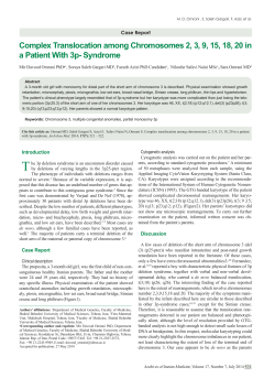
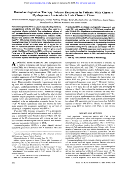
American Journal of Biomedical and Life Sciences
2014; 2(2): 46-54
Published online May 30, 2014 (http://www.sciencepublishinggroup.com/j/ajbls)
doi: 10.11648/j.ajbls.20140202.13
Cytogenetic investigation in prenatal specimens for
effective prognosis of pregnancy related complications
Binay Kumar Raut1, Lakshman Kumar Balasubramanian2, Mukesh Kumar Jha3,
Shyam Sundar Malla1, Moka Rajasekhar4
1
Department of Biochemistry, Kathmandu University School of Medical Sciences, Dhulikhel, Kavre, Nepal
Department of Biotechnology, Kongunadu Arts and Science College, Coimbatore, Tamilnadu, India
3
Department of Physiology, Kathmandu University School of Medical Sciences, Dhulikhel, Kavre, Nepal
4
Department of Biotechnology, School of Life Sciences, Manipal University, Manipal, Karnataka, India
2
Email address:
[email protected] (B. K. Raut), [email protected] (L. K. Balasubramanian), [email protected] (M. K. Jha),
[email protected] (S. S. Malla), [email protected] (M. Rajasekhar)
To cite this article:
Binay Kumar Raut, Lakshman Kumar Balasubramanian, Mukesh Kumar Jha, Shyam Sundar Malla, Moka Rajasekhar. Cytogenetic
Investigation in Prenatal Specimecimens for Effective Prognosis of Pregnancy Related Complications. American Journal of Biomedical
and Life Sciences. Vol. 2, No. 2, 2014, pp. 46-54. doi: 10.11648/j.ajbls.20140202.13
Abstract: Background: Alterations in chromosomal content of mother and infant are central characteristics of various
complications related to pregnancy and early childhood. About 60% of the pregnancy losses, 2-3% of all the neonates and
50% of childhood deafness, blindness, mental retardation and 1 to 10 % of all the malignancies are directly due to genetic
factors. Hence cytogenetic testing of pre and post natal samples can prove to be useful for discovery of non-invasive
markers for prevention of such conditions beforehand. Aim of the study: The present study was carried out to detect
numerical and structural abnormalities in 56 subjects with repeated miscarriages, bad obstetric history and sub fertility by
analysing peripheral blood, products of conception (POC) material, and recovered cell lines from prenatal samples.
Methods: Conventional cytogenetics: Peripheral blood culture (PBC) supplemented with mitogen Phytohemagglutinin
(PHA), metaphase chromosomes was harvested after 72 hours for chromosome analysis. Tissue cytogenetics: Culture of
solid tissue was used as a source for mitotic cells from products of conception (POC) from first trimester spontaneous
abortions for aneuploidy detection; and Prenatal chromosome analysis was performed by either chorionic villus sampling
(CVS), amniotic fluid and cord blood after culture. Image acquisition and analysis was performed by using automated
karyotyping (IKAROS) software based on G, C and R banding. Results: Chromosomal abnormalities were located in all
types of specimens but were predominantly observed in recurrent pregnancy loss (RPL) and product of conception (POC)
samples. Aberrations observed were mainly translocations, satellites, additions in RPL cases like 46,XX with instances of
(D/D,D/G,G/Gassociations);45,XX,rob(13;14);46,XXt(4;21);46,XX,(9qh+);46,XX,(14ps+);46,XX, t(5;6) and ploidy
involving 67,XX+;64,XXX+;69,XXX;63,XXX;58,XX+ in the POC cases. Conclusion: Cytogenetic screening could
provide to be a useful method for monitoring patients with abnormal pregnancies. The cytogenetic result is an independent
prognostic indicator, with certain karyotypes associated with a good prognosis for the better treatment.
Keywords: Products of Conception (POC), Peripheral Blood Culture (PBC), Karyotyping (G, C and R Banding)
1. Background and Introduction
All species are affected by genetic diseases.
Chromosomal manifestations are diverse and numerous,
including early embryonic death, minor to major congenital
defects, and infertility or sterility. Pregnancy loss is defined
as the unexpected and unplanned spontaneous loss of
pregnancy before the fetus becomes capable of extra
uterine survival.
About 15% of all recognized pregnancies end in
spontaneous abortions. The single most common reason is
the presence of a chromosome abnormality in the fetus
particularly if the loss occurs early in the pregnancy.
Approximately 50% of such miscarriages are associated
with cytogenetic abnormalities, with trisomy being the most
frequent, followed by polyploidy and monosomy X
[Kalousek et al., 1993]. Such miscarriages are thought to
American Journal of Biomedical and Life Sciences 2014; 2(2): 46-54
occur on a random basis, with an increasing frequency of
trisomy with advancing maternal age [Hassold and Chiu,
1985]. More than 99% of chromosomally abnormal
pregnancies result in miscarriage, most of which occur
prior to 10 weeks gestation [Jacobs and Hassold, 1987].
About 60% of recurrent miscarriages might be caused by
chromosomal aberrations in the embryo [Carp et al., 2004].
About 3-5% of couples with recurrent miscarriage have one
partner with balanced chromosomal rearrangement and
carriers of reciprocal translocations are thought to have
higher frequencies [Fred Kavalier 2005; Ogasawara et al.,
2004].
With respect to cytogenetic studies in the above field, the
most frequently applied human tissues are peripheral blood,
amniotic fluid, chorionic tissue and skin fibroblasts.
Amniocytes and chorionic cells are important in prenatal
diagnostics [Eisenberg and Wapner 2002], and 1–15 days
of cell culture are needed to obtain metaphase cells in this
case [Wegner 1999]. Often, the cytogenetic results provide
for definitive diagnosis and monitoring strategies.
The recent developments in molecular cytogenetic
technologies, in association with conventional cytogenetic
analysis, have improved the accuracy of the results and led
to the finding of new chromosomal abnormalities. Hence a
broad base of knowledge is necessary in order to
understand, diagnose and advice patients about the complex
field of pregnancy related complications.
2. Aim of the Study
The main aim of the present study was to perform
cytogenetic analysis to detect numerical and structural
abnormalities by using automated karyotyping (IKAROS)
software in repeated miscarriage samples including
products of conception (POC) material and recovered cell
lines from prenatal samples
3. Materials and Methods
The present study was carried out to detect numerical
and structural chromosomal abnormalities in 56 subjects
(42 patients and 14 prenatal samples).
3.1. Conventional Cytogenetics
Peripheral blood culture (PBC) supplemented with
mitogen Phytohemagglutinin (PHA) was setup and
metaphase chromosomes were harvested after 72 hours for
chromosome analysis after arrest by colchicines.
3.2. Tissue Cytogenetics
Culture of solid tissue was used as a source for mitotic
cells from products of conception (POC) from first
trimester spontaneous abortions for aneuploidy detection
After receiving the sample in Sodium saline, a small
piece of the tissue was taken in a petridish. The tissue was
washed with PBS at least 4 to 5 times and placed on a six
47
well plate and supplemented with 3ml of DMEM with 10%
FBS. The tissues were grown at 37oC in CO2 incubator and
later transferred to a T25 flask. When the flask was 100%
confluent, culture was trypsinized. After 48 hours
incubation, cells were harvested. Metaphase cells were
studied for the abnormalities.
Prenatal chromosome analysis was performed by
amniotic fluid, chorionic villus sampling (CVS) and cord
blood after culture.
The amniotic fluid collected was spun at 800 rpm for 10
minutes. The pellet was suspendended in 5 ml of growth
medium (DMEM). The primary culture was setup i.e., the
collected pellet in 3ml of Amniomax medium which
contains 15% FBS, and distributed in three T25 flasks
For CVS and cord blood, the tissue culture method and
blood culture method mentioned above was used
3.3. High Resolution Prometaphase Chromosome
Preparation
In addition to routine chromosome culture, Methotrexate,
Thymidine and Ethidium bromide were added to cultures
for culture and harvest for high-resolution prometaphase
chromosomes [Yunis et al. 1976]. Briefly, on the day prior
to harvest was added 50µl of Methotrexate to block the
DNA replication followed by incubation for another 16
hours. The next day was added 50µl of Thymidine and
culture further incubated for another 3 hours. Finally 50µl
of Ethidium bromide solution was added and culture
incubated for one hour for excessive chromosome
condensation. Then 50µl colcemid solution was added to
culture, incubated for 30 minutes and harvested and studied.
G,R and C banding techniques were performed as per the
standard protocols and stained slides were screened for
good metaphase spreads and the metaphases were captured
under 100X oil immersion of the microscope (Ziess)
attached with CCD camera. 50 metaphases were captured
per sample and the karyotypes were analyzed with the help
of Metasystem Ikaros software to detect any numerical or
structural chromosomal abnormalities.
4. Results
Table 1 and 2 show the clinical data associated with the
present study.The study included 42 patients who presented
with repeated pregnancy loss, Bad obstetric history,
subfertility and 14 products of conception and amniotic
fluid samples. For the latter, recovered cell lines from
miscarriage material and marker test positive samples were
analysed (Figure 1).
It was found from the clinical data that the age groups of
the patients were between 25-35 years.
Chromosomal abnormalities were located in all types of
specimens using the three banding techniques namely G,R
and C banding(Figure 2) but were predominantly observed
in recurrent pregnancy loss (RPL) and product of
conception (POC) samples(Figure 3). Aberrations observed
were mainly translocations, satellites, additions in RPL
48
Binay Kumar Raut et al.: Cytogenetic Investigation in Prenatal Specimens for Effective Prognosis of
Pregnancy Related Complications
cases like 46,XX with instances of (D/D,D/G,G/G
associations)(Figure 4); 46,XX,(15ps+)(Figure 5);46,XX,
t(5;6)
(Figure
6)45,XX,rob(13;14)(Figure
7);46,XXt(4;21);46,XX,(9qh+).46 XX (Figure 8) was
predominant in amniotic fluid whereas Monosomy(Figure
9
)
and
ploidy
involving
67,XX+;64,XXX+;69,XXX(Figure
10) ;63,XXX;58,XX+ ,81 XXYY (Figure 11) were
observed in the POC cases in addition to normal karyotypes.
Table 1. Cytogenetics data of Repeated pregnancy loss, Bad Obstetric
History and subfertility patients
Age /
Sex
Clinical diagnosis
1.
28/F
Repeated Pregnancy
Loss (RPL)
46,XX
2.
28/F
RPL
46,XX
3.
27/F
RPL
46,XX
4.
33/F
RPL
5.
32/F
6.
30/F
7.
25/F
RPL
8.
25/F
9.
S.No
S.No
Age /
Sex
Clinical diagnosis
Results
37.
25/F
RPL
46,XX
38.
28/F
BOH
46,XX
39.
23/F
RPL
46,XX,t(5;6)
40.
25/F
RPL
46,XX
41.
28/F
RPL
46,XX
42.
30/F
RPL
46,XX
Table 2. Cytogenetic analysis for recovered cell lines from amniotic fluid,
Products of conception of triple marker positive and miscarriage patients
respectively
Results
Cell lines and Initial
Karyotype
Recovered cell lines
Karyotype
1.
45,X
67,XX+
POC
2.
46,XY
46,XY
POC
46,XX
3.
46,XY
46,XY
POC
BOH
46,XX
4.
46,XX
64,XXX+
POC
BOH
5.
46,XY
46,XY
POC
6.
46,XX
46,XX
POC
7.
69,XXX
69,XXX
POC
RPL
46,XX
46,XX, Instances of
(D/D,D/G,G/G
associations)
46,XX
28/F
RPL
46,XX
8.
46,XX/47,XY+16
63,XXX
POC
10.
36/F
RPL
46,XX
9.
46,XX, (15ps+)
58,XX+
POC
11.
25/F
RPL
46,XX
10.
46,XX
46,XX
AF
12.
25/F
RPL
46,XX
11.
46,XY
46,XY
AF
13.
25/F
RPL
46,XX
14.
28/F
46,XX
12.
46,XX
46,XX
AF
13.
46,XX
46,XX
AF
15.
25/F
14.
46,XX
46,XX
AF
16.
28/F
RPL
One miscarriage and
infertility
BOH
17.
30/F
RPL
46,XX
18.
27/F
RPL
46,XX
19.
29/F
BOH
46,XX
20.
20/F
BOH
46,XX
21.
26/F
RPL
46,XX
22.
33/F
RPL
46,XX
23.
27/F
BOH
46,XX
24.
23/F
RPL
46,XX
25.
28/F
RPL
46,XX
26.
23/F
RPL
45,XX,rob(13;14)
27.
27/F
RPL
46,XX
28.
28/F
RPL
46,XX
29.
30/F
RPL
46,XX,t(4;21)
30.
23/F
RPL
46,XX
31.
27/F
RPL
46,XX
32.
28/F
RPL
46,XX
33.
29/F
RPL
46,XX
34.
25/F
RPL
46,XX
35.
26/F
RPL
46,XX, (9qh+)
36.
26/F
RPL
46,XX,(14ps+)
S.No
Source
45,XX,rob(13;14)
46,XX, (15ps+)
Figure 1. Recovered cell lines from amniotic fluid sample (Fibroblast cells)
American Journal of Biomedical and Life Sciences 2014; 2(2): 46-54
Figure 2. Different banding techniques G banding, R banding and C banding
Figure 3. A placental tissue from products of conception - Endoreduplication
Figure 4. D/D, D/G and G/G associations (H/o repeated miscarriages)
49
50
Binay Kumar Raut et al.: Cytogenetic Investigation in Prenatal Specimens for Effective Prognosis of
Pregnancy Related Complications
Figure 5. 46,XX,(15ps+) in a female with Bad Obstetric History
Figure 6. Karyotype 46, XX.t (5;6) – in a patient with repeated miscarriages
Figure 7. 45, XX, rob (13;14)) – in patient with missed abortion and subsequent infertility
American Journal of Biomedical and Life Sciences 2014; 2(2): 46-54
Figure 8. 46,XX karyotype (Amniotic fluid culture)
Figure 9. Monosomy (45, X) in missed aborted material (POC)
Figure 10. Triploidy (Products of conception sample)
51
52
Binay Kumar Raut et al.: Cytogenetic Investigation in Prenatal Specimens for Effective Prognosis of
Pregnancy Related Complications
Figure 11. Tetraploidy (Products of conception sample)
5. Conclusion
Here we report different numerical and structural
abnormalities in different prenatal specimens which could
be further probed and investigated to serve as markers for
pregnancy related complications. Hence cytogenetic
screening could provide to be a useful method for
monitoring patients with abnormal pregnancies. The
cytogenetic result is an independent prognostic indicator,
with certain karyotypes associated with a good prognosis
for the better treatment.
Early identification of the possible cause of fetal loss can
significantly reduce long term psychological distress in
women with miscarriages and enables improved genetic
counseling for those couples in future pregnancies.
References
[7]
Bagby, G. C., Lipton, J. M., Sloand, E. M., and Schiffer, C.
A. (2003).Marrow failure. J. Med. Genet., 40, 1-10.
[8]
Bennett JM, Catovsky D, Daniel MT, Flandrin G, Galton
DA, Sultan C. (1952) Proposals for the classification of the
myeloidysplastic syndrome. Br J Hematol 1952; 51: 189199
[9]
Bloom D (1954) Congenital telangeetatic erythema
resembling lupus erythematosus in dwarfs. Am J. Dis child
88; 754-758.
[10] Brady K, Duff P, Harlass FE, Reid S (1991). Role of
amniotic fluid cytogenetic analysis in the evaluation of
recent fetal death Am J Perinatol;8(1):68-70.
[11] Carp H, Feldman B, Oelsner G, Schiff E. (2004) Parental
karyotype and subsequent live births in recurrent
miscarriage. Fertil Steril 81; 1296-1301.
[12] Carp H, Toder V, Aviram A, Daniely M, Mashiach S and
Barkai G (2001) Karyotype of the abortus in recurrent
miscarriage. Fertil Steril 75,678–682.
[1]
Alter BP. Clinical features of Fanconi’s anaemia. In: Young
NS, Alter BP, eds. Aplastic anemia: acquired and inherited.
Philadelphia: Saunders, 1994:275-308.
[13] Carrera M (2001) Screening prenatal de aneuploidı´as: QFPCR y FISH. Prog Diag Prenat 13,262–266.
[2]
Amor-Gueret M. Bloom’s syndrome. Orphanet encyclopedia.
[cited on 2004. Available from: ttp//www.orpha.net/.
[14] Caspersson T, Zech L, Johansson C: Differential binding of
alkylating fluorochromes in human chromosomes. Exp Cell
Res 1970, 60:315-319.
[3]
Andaloussi El and Bilhou-Nabera C (2007) Case Report
New Complex Chromosomal Translocation in Chronic
Myeloid
Leukaemia: t(9;18;22)(q34;p11;q11)
J.
Biomedicine and Biotechnology Vol 2007, Article ID 92385,
3 pages.
[4]
[5]
[6]
Arrighi FE and Hsu TE (1971) Localization of
heterochromatic regions of human chromosomes.
Cytogenetics 10; 81-86.
Auerbach, A D., Adler, B., and Chaganti, R. S. K. (1981).
Prenatal and postnatal diagnosis and carrier detection of
Fanconi anemia by the cytogenetic method. Pediatrics, 67,
128-35.
Auerbach, A. D., Buchwald, M., and Joenje, H. (2001).
Fanconi anemia. In: The Metabolic and Molecular Bases of
Inherited Disease,(Eds. CR Scriver, WS Sly, B Childs, et al.),
1, 753-68 McGraw-Hill, New York.
[15] Chandley AC (1983). Infertility and recurrent abortion In.
Emery AEH, Rimoin (Eds). Principles and practice of
Medical genetics, Churchill Livingston, New York.
[16] de Braekeleer M (1987) “Variant Philadelphia translocations
in chronic myeloid leukemia,” Cytogenetics and Cell
Genetics, vol. 44, no. 4, pp. 215–222, 1987.
[17] Dicken CH, Dewald G, and Gordon H (1978) Sister
chromatid exchanges in Bloom syndrome. Arch Dermatol
114(5); 755-760.
[18] Dutrillaux B (1973) Nouveau systeme de marquage
Chromosomeiqe les bandes T. Chromosoma 41; 395;402.
[19] Eisenberg B, Wapner RJ (2002) Clinical procedures in
prenatal diagnosis. Best Pract Res Clin Obstet Gynaecol
16:611–627.
American Journal of Biomedical and Life Sciences 2014; 2(2): 46-54
[20] Ellis NA, German J. Molecular genetics of Bloom’s
syndrome. Hum Mol Genet 1996;5:1457-63.
[21] Fred Kavalier (2005) Investigation of recurrent miscarriages
BMJ. 2005 July 16; 331(7509): 121–122.
[22] Fryns JP, Kleczkowska A, Kubien E, Petit P, Vanberghe H
(1984) Cytogenetic survey in couples with recurrent fatal
wastage. Hum Genet 65;336-354.
[23] German J and Crippa LP (1966) chromosomal breakage in
diploid cell lines from Bloom’s syndrome and Fanconi’s
anemia. Ann Genet 9: 143-154.
[24] Goodpasture C Bloom SE, Hsu TC and Arrighi FE (1975)
Human nucleolus organizers: the satellites or the stalks? Am
J Hum Genet 28;;559-566.
[25] Hassold, T. and Chiu, D. (1985) Maternal age specific rates
of numerical chromosome abnormalities with special
reference to trisomy. Hum. Genet., 70, 11–17.
[26] Hatice Ilgin, Ayse Nurten Akarsu, Fatma Isik Bökesoy (1999)
Cytogenetic and Phenotypic Findings in Turkish Patients
With Fanconi’s Anemia Tr. J. of Medical Sciences 29; 151154
[27] Horrike S, Taniwaki M, Misawa S, Abe T (1988)
Chromosome abnormalities and karyotypic evolution 83
patients with myelodysplastic syndrome and predictive
value for prognosis. Cancer 62: 1129-38.
[28] Hossein Mozdarani, Anahita Mohseni Meybodi, Shabnam
Zari-Moradi (2008) A cytogenetic study of couples with
recurrent spontaneous abortions and infertile patients with
recurrent IVF/ICSI failure. Indian J of Hum Genet 14(1) 1-6.
[29] Hsu, T. C. Mammalian chromosomes in vitro, I. Karyotype
of man. Journal of Heredity 43, 167–172 (1952).
[30] Jacobs P.A. and Hassold T.J. (1987) Chromosome
abnormalities: origin and etiology in abortions and live
births. In Vogal, F. and Sperling, K. (eds), Human Genetics.
Springer-Verlag, Berlin, pp. 233–244.
[31] Jacobs RH, Cornbleet MA, Vardiman JW, Larson RA, Le
Beau MM, Rowley JD(1986) Prognostic implications of
morphology and karyotype in primary myelodysplastic
syndromes. Blood 67:1765-72.
[32] Jemal A, Thomas A, Murray T, Thun M (2002). "Cancer
statistics, 2002". CA Cancer J Clin 52 (1): 23–47.
[33] Jim Martin Southern Ontario Fertility Technologies
(S.O.F.T.) 555 Southdale Rd., E., Suite 107, London,
Ontario, N6E 1A2.
[34] Joenje H, Oostra AB, Wijker M, Di SFM, Van BCG,
Rooimans MA, Ebell W, Van WM, Pronk JC, Buchwald M,
Arwert F (1997). Evidence for at least eight Fanconi anemia
genes. Am J Hum Genet;61:940-944.
[35] Kadam PR, Nangangund GJ, Advani SH (1990), The
occurrence of variant Ph translocation in chronic myeloid
leukemia (CML); a report of 6 cases. Hematol Oncol 8;
303-312.
[36] Kalousek, D.K., Pantzar, T., Tsai, M. et al. (1993) Early
spontaneous abortion: morphologic and karyotypic findings
in 3,912 cases. Birth Defects, 29, 53–61.
53
[37] Kim JM, Sim AS, Lee EH (2006) Amniotic chromosome
analysis in pregnant women identified by triple marker
testing as screens positive. Korean J Lab Med 26(2); 12330.
[38] Lerardi-Curto L (2009) Chromosomal breakage syndromes.
eMedicine Pediatrics, Webmed.
[39] Marquard K, Westphal LM, Milki AA and Lathi RB (2009)
Etiology of recurrent pregnancy loss in women over the age
of 35 years. Fertil Steril 30th July 2009.
[40] Mitelman F (1993), “The cytogenetic scenario of chronic
myeloid leukemia,” Leukemia and Lymphoma, vol. 11, pp.
11–15, 1993.
[41] Mitelman F, Johansson B, and Mertens F (2006). Eds,
“Mitelman Database of Chromosome Aberrations in
Cancer,” http://cgap .nci.nih.gov/Chromosomes/Mitelman,
August 2006.
[42] Moorhead, P.S., Nowell, P.C., Mellman, W.J., Battips, D.M.
and Hungereford, D.A. (1960). Chromosome preparations of
leukocytes cultured from human peripheral blood. Exp. Cell
Res. 20: 613-616.
[43] Naeimeh Tayebi, Hossain Khodaei (2008) Bloom’s
syndrome in a 12-year-old Iranian girl Ind J. Hum Genet
14(3); 103-105.
[44] Nowell, P., & Hungerford, D. A minute chromosome in
human chronic granulocytic leukemia. Science 132, 1497
(1960).
[45] Ogasawara M, Ozaki Y, Sato T, Suzumori N, Suzumori K
(2004) Poor prognosis of recurrent aborters with either
maternal or paternal reciprocal translocations. Fertil Steril
81;367-373.
[46] Reid DE, Ryan KJ, Benirsche K. (1972) Principles and
management of human reproduction, Saunders, Philadelphia,
London, Toronto.
[47] Rowley JD (1973) “A new consistent chromosomal
abnormality in chronic myelogenous leukaemia identified
by quinacrine fluorescence and Giemsa staining,” Nature,
vol. 243, no. 5405, pp.290–293, 1973.
[48] Sandberg A. A (1980) The Chromosomes in Human Cancer
and Leukemia, Elsevier North Holland, New York, NY,
USA, 1980.
[49] Sanja cirkovic, Marija Guc-Scekic, Dragana Vujic and D
micic (2006) Cytogenetic diepoxybutane sensitivity in
Serbian children with Fanconi anemia. Arch Biol Sci
Belgrade. 58 (4); 215-219.
[50] Santalo J, Catala V and Badenas J (1987) Chromosomal
abnormalities and IVF. In Egozcue J (ed.) Cellular Aspects
of In Vitro Fertilization. Cell Biology Reviews. Leiola, pp.
63–72.
[51] Schoch C and Haferlach T (2007) Cytogenetics in acute
myeloid leukemia. Current Oncology Report vol. 4(5) 390391.
[52] Schroeder TM, Tilgen D, Kruger J, Vogel F. Formal genetics
of Fanconi’s anemia. Hum Genet 1976;32:257-88.
[53] Seabright M (1971) A rapid banding technique for human
chromosomes. Lancet, 7731:971-972.
54
Binay Kumar Raut et al.: Cytogenetic Investigation in Prenatal Specimens for Effective Prognosis of
Pregnancy Related Complications
[54] Shaffer LG, Tommerup N, ISCN 2005: An international
system for cytogenetic nomenclature. S. Karger, Basel,
Switzerland 2005.
[55] Shahrabani-Gargir L, Shomrat R, Yaron Y, Orr-Urtreger A,
Groden J, Legum C. High frequency of a common bloom
syndrome Ashkenazi mutation among Jews of polish origin
Genet Test 1998;2:293-6.
[56] Shalev E, Zalel Y, Weiner E, Cohen H, Shneur Y (1994) The
role of cordocentesis in assessment of mosaicism found in
amniotic fluid cell culture. Acta Obstet Gynecol Scand;
73(2):119-22.
[57] Tjio, J. H., & Levan, A. The chromosome number of man,
Hereditas 42, 1–6 (1956)
[58] Toogeh Gl, Najafi AH, and Keyhani M (2003) Cytogenetic
findings in acute myeloid leukemia. Acta Medica Iranica 41
(4) 227-232.
[59] Verma RS, Babu A (1994) Human chromosomes: principles
and techniques, 2nd edn McGraw-Hill, New York.
[60] Vundinti BR, Kerketta L, Jijina F and Ghosh K (2009)
Cytogenetic study of myelodysplastic syndrome from India.
Indian J Med Res 130: 155-159.
[61] Warburton D, Stein Z, Kline J, and Susser M, et al. (1980)
Chromosome abnormalities in spontaneous abortion: data
from the New York City study. In Porter IH and Hook EB
(eds) Human Embryonic and Fetal Death. Academic Press,
New York, pp. 261–287.
[62] Wegner RD (1999) Diagnostic cytogenetics. Springer,
Berlin.
[63] Yunis JJ: High resolution of human chromosomes. Science
1976, 191:1268-1270.
© Copyright 2026 ExpyDoc