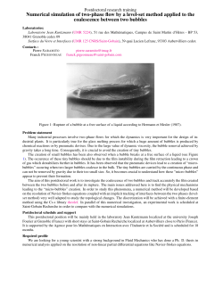
DISEASES OF AQUATIC ORGANISMS
Dis Aquat Org
Vol. 111: 191–205, 2014
doi: 10.3354/dao02790
Published October 16
FREE
ACCESS
Decompression sickness (‘the bends’) in sea turtles
D. García-Párraga1,*, J. L. Crespo-Picazo1, 2, Y. Bernaldo de Quirós3, V. Cervera4,
L. Martí-Bonmati5, J. Díaz-Delgado3, M. Arbelo3, M. J. Moore6, P. D. Jepson7,
Antonio Fernández3
1
Oceanografic, Veterinary Services, Parques Reunidos Valencia, Ciudad de las Artes y las Ciencias,
C/ Eduardo Primo Yúfera 1B, 46013 Valencia, Spain
2
VISAVET Center and Animal Health Department, Veterinary School, Complutense University of Madrid,
Av Puerta del Hierro s/n, 28040 Madrid, Spain
3
University of Las Palmas de Gran Canaria, Institue of Animal Health, C/Transmotaña s/n, Arucas, 35416, Las Palmas, Spain
4
Hospital Veterinario Valencia Sur, Avda. Picassent, 28, 46460 Silla, Valencia, Spain
5
Grupo de Investigación Biomédica en Imagen GIBI230, Radiology Department, Hospital Universitario y Politécnico La Fe,
Av. Bulevar Sur s/n, 46026 Valencia, Spain
6
Woods Hole Oceanographic Institution, Department of Biology, 266 Woods Hole Road, Woods Hole, MA 02543, USA
7
Institute of Zoology, Zoological Society of London, Regent’s Park, London, NW1 4RY, UK
ABSTRACT: Decompression sickness (DCS), as clinically diagnosed by reversal of symptoms with
recompression, has never been reported in aquatic breath-hold diving vertebrates despite the
occurrence of tissue gas tensions sufficient for bubble formation and injury in terrestrial animals.
Similarly to diving mammals, sea turtles manage gas exchange and decompression through
anatomical, physiological, and behavioral adaptations. In the former group, DCS-like lesions have
been observed on necropsies following behavioral disturbance such as high-powered acoustic
sources (e.g. active sonar) and in bycaught animals. In sea turtles, in spite of abundant literature
on diving physiology and bycatch interference, this is the first report of DCS-like symptoms and
lesions. We diagnosed a clinico-pathological condition consistent with DCS in 29 gas-embolized
loggerhead sea turtles Caretta caretta from a sample of 67. Fifty-nine were recovered alive and 8
had recently died following bycatch in trawls and gillnets of local fisheries from the east coast of
Spain. Gas embolization and distribution in vital organs were evaluated through conventional
radiography, computed tomography, and ultrasound. Additionally, positive response following
repressurization was clinically observed in 2 live affected turtles. Gas embolism was also observed
postmortem in carcasses and tissues as described in cetaceans and human divers. Compositional
gas analysis of intravascular bubbles was consistent with DCS. Definitive diagnosis of DCS in sea
turtles opens a new era for research in sea turtle diving physiology, conservation, and bycatch
impact mitigation, as well as for comparative studies in other air-breathing marine vertebrates and
human divers.
KEY WORDS: Gas bubbles · DCS · Caretta caretta · Loggerheads · Bycatch · Hyperbaric treatment ·
Gas embolism · Breath-hold divers
Resale or republication not permitted without written consent of the publisher
Decompression sickness (DCS) is a clinical diagnosis encompassing a wide range of manifestations
related to formation of gas bubbles within supersatu-
rated tissues after decompression (Barratt et al. 2002,
Francis & Mitchell 2003). In human divers, the effects
range from trivial to fatal, and most often involve
neurological and musculoskeletal symptoms (Francis
& Simon 2003, Vann et al. 2011), including severe
*Corresponding author: [email protected]
© Inter-Research 2014 · www.int-res.com
INTRODUCTION
192
Dis Aquat Org 111: 191–205, 2014
pain. In an analysis of 1070 central nervous system
DCS cases, 77% involved the spinal cord (Francis et
al. 1988). A wide range of symptoms are caused
directly or secondarily by the mechanical, embolic,
and biochemical effects of intra- and extravascular
bubbles (Vann et al. 2011). Direct effects include the
distortion of tissues and vascular obstructions, while
secondary effects include endothelial damage, capillary leakage, plasma extravasation, and hemoconcentration (Vann et al. 2011). Definitive diagnosis of
DCS is difficult and only confirmed by successful
recompression treatment in a hyperbaric chamber
(Ferrigno & Lundgren 2003).
Breath-hold diving vertebrates, including marine
mammals and sea turtles, classically have been considered to be protected against DCS through
anatomical, physiological, and behavioral adaptations (Berkson 1967, Rothschild & Martin 1987,
Burggren 1988, Lutcavage & Lutz 1997, Piantadosi
& Thalmann 2004, Fossette et al. 2010, Castellini
2012). However, an acute and systemic gas and fat
embolic syndrome similar to DCS in human divers
was described in beaked whales that stranded in
temporal and spatial association with military exercises involving high-powered sonar (Jepson et al.
2003, Fernández et al. 2005). Since this first report,
there has been accumulating evidence demonstrating the presence of gas bubbles in diving marine
mammals (Jepson et al. 2005, Moore et al. 2009,
Bernaldo de Quirós et al. 2012, Dennison et al.
2012), including dysbaric osteonecrosis (Moore &
Early 2004) and gas embolism in bycaught animals
(Moore et al. 2009). Although these findings have
challenged our understanding of diving physiology
in these species, conclusive clinical data (i.e. diagnosis and therapy) supporting the occurrence of
DCS are lacking due to the complexity of working
with wild marine mammals.
Sea turtles are among the longest- and deepestdiving air-breathing marine vertebrates (Byles 1988,
Sakamoto et al. 1990, Houghton et al. 2008). They
may spend over 90% of their life-time submerged in
apnea (Lutcavage & Lutz 1997) and efficiently use
oxygen through cardiovascular adjustments, similar
to other air-breathing vertebrates (Rothschild & Martin 1987, Burggren 1988, Southwood et al. 1999,
Southwood 2013). In addition, although osteonecrosis-type lesions (among the few long-term lesions
observable after certain episodes of DCS) have been
described in mosasaurs and sea turtle fossils from the
Cretaceous Age, such lesions are very rarely described
in marine reptile species younger than the Miocene
Age (Rothschild & Martin 1987). This suggests that
more recent taxa have evolved physiological and
behavioral adaptations to mitigate hyperbaric conditions such as DCS.
Bycatch is a well-documented, worldwide problem
resulting in considerable mortality of non-targeted
species (Lewison et al. 2004a). Over recent decades,
there has been a dramatic global decline in sea turtle
populations, with 6 of 7 species currently categorized as Vulnerable, Endangered, or Critically Endangered on the IUCN Red List (www.iucnredlist.
org; accessed 14 January 2014). Fishery bycatch is
recognized as the greatest threat to their conservation (Wallace et al. 2010) and is considered a moderate or high threat for more than 75% of all sea turtle
Regional Management Units globally (Wallace et al.
2011, Lewison et al. 2013). Approximately 85 000 sea
turtles were reported incidentally captured worldwide from 1990 through 2008, but true total bycatch
is estimated to be at least 2 orders of magnitude
higher (Wallace et al. 2010). Total numbers of global
bycaught sea turtles (Lewison et al. 2004b, Hamann
et al. 2010, Wallace et al. 2010) and resulting mortality (Lutcavage & Lutz 1997, Epperly et al. 2002,
Hamann et al. 2010) remain unclear.
The primary limitation in bycatch estimates is the
lack of reliable comprehensive information on total
fisheries effort, bycatch in small-scale fisheries (Wallace et al. 2010, Casale 2011), and the rate of survivorship of released animals (Chaloupka et al. 2004,
Mangel et al. 2011). The rate of survivorship following interaction is considered to be one of the main
obstacles to understanding the true impact of fisheries on sea turtle populations (Lewison et al. 2013).
Consideration of causes of sea turtle mortality resulting from fisheries interactions has largely focused on
the effects of drowning and direct trauma from gear
(Poiner & Harris 1996, Gerosa & Casale 1999, Casale
2011, Lewison et al. 2013). The present work describes a previously undescribed condition that can
compromise post-release survivorship of incidentally
captured sea turtles.
In this study, 67 loggerhead turtles (59 alive, 8
dead) bycaught in trawls and gillnets at depths ranging from 10 to 75 m, were evaluated by intensive clinical and pathological examination. Gas embolism
(GE) was a consistent finding in a large proportion of
live and dead animals. Clinical signs, diagnostic
imaging, gross and histological observations, and
response to recompression and controlled decompression treatment strongly suggest that marine airbreathing vertebrates can suffer from DCS. These
findings offer a new paradigm to consider in many
different aspects of sea turtle research, conservation,
García-Párraga et al.: ‘The bends’ in sea turtles
and management, including basic patho-physiological aspects of diving adaptations, implications on
post-capture survivorship estimates, bycatch impact
mitigation strategies and devices, and clinical treatment of affected turtles, as well as potential additional risks associated with intentional capture of
diving turtles.
MATERIALS AND METHODS
Animal acquisition
All sea turtles included in this project were under
the authority of the ‘Consellería de Infraestructuras,
Territorio y Medio Ambiente’ of the Valencia Community Regional Government in collaborative official
agreement with the Oceanografic Aquarium of the
‘Ciudad de las Artes y las Ciencias of Valencia’ for
animal rehabilitation and posterior release, and for
the postmortem examination of dead individuals.
In 2011, an active campaign involving fishermen
from the Valencian coast of Spain was initiated to
collect all (live and dead) sea turtles incidentally captured by gillnets and trawling so that bycaught animals could be medically evaluated. During the
period from 1 January 2011 to 2 January 2014, a total
of 67 bycaught loggerhead turtles Caretta caretta
were received. Eleven turtles arrived dead and 56
arrived alive, although 5 of the 56 live turtles died
within 72 h. All live animals received comprehensive
clinical examination. Examination of all dead turtles
included necropsy and histopathology.
For all cases, the date of capture, fishing depth, and
sea surface temperature at the originating port were
documented (SeaTemperature: www.seatemperature.org; accessed 14 January 2014). Any comments
from fishermen related to the condition and behavior
of turtles upon capture were also noted.
Clinical diagnosis
All live bycaught turtles were examined within the
first 24 h (average 12 h). Evaluation included routine
general veterinary physical and neurological examination, hematology, and biochemistry, followed by
imaging studies.
Blood was collected from the dorsal cervical sinus
with a 5 ml syringe and 21G 40 mm hypodermic
needle (Henry Schein) and transferred to 2 ml
lithium heparin tubes (Aquisel®) for immediate
analysis (maximum elapsed time of 1 h). Analysis
193
included automated hematology with an Abbott
Celldyn 3700SL hemocytometer (Abbott Laboratories), standard manual hematocrit determination and
cytological study including manual differential count,
and complete biochemistry and electrolyte panel using
an Olympus AU400 autoanalyzer (Mishima Olympus).
Diagnostic imaging studies included the following:
(1) Plain radiographic evaluation with a Philips
Practix 400 unit (Philips Medical Systems) and a Kodak Direct View Classic CR System (Carestream
Health) with 35 × 43 cm Kodak cassettes (Kodak PQ
Storage Phosphor Screen Regular and 100 Microns,
Carestream Health) in dorsal-ventral (DV), cranialcaudal (CC), and lateral-lateral (LL) projections. Focal
distances varied between 1 and 1.5 m, using average
exposure values between 75 and 120 kV and 7.2 to
20 mAs depending on projections and animal size.
Digital images were processed afterwards with Kodak
Acquisition Software (Onyx-RAD Diagnostic Viewer)
for better visualization and image interpretation. Some
dead bycaught turtles were also radiographed.
(2) Ultrasonographic general examination was conducted using a General Electric Logiq E Vet ultrasound machine with commercial linear, phase-array,
and microconvex probes (models 12LRS [GE Healthcare], 3S [GE Medical Systems], and 8CRS [GE Medical Systems], respectively).
(3) Selected individuals with DCS compatible signs
were examined by computed tomography (CT) using
a Toshiba Aquilion 16 CT unit (Toshiba Medical Systems). Acquisition parameters through whole-body
exploration of the turtle were 5 mm slice thickness
and 5 mm slice interval, with 0.5 mm retro-recon
acquisition under lung and mediastinal algorithms.
Images were post-processed with Osirix software
version 3.3.1 (Pixmeo) and Philips Brilliance Workspace CT software (Koninklijke Philips). A 3D air volume was recreated through volumetric segmented
reconstruction (volume rendering).
Based on imaging findings upon arrival at the
rehabilitation center and/or postmortem examinations, the severity of gas embolism was scored based
on the total amount of intravascular gas observed
and its distribution, as follows (see Table 1):
(1) Mild embolism: a small amount of gas was only
evident in the kidney region on ultrasound and LL
radiographic projection.
(2) Moderate embolism: a larger volume of gas was
present in the kidney region, being clearly evident in
ultrasound, LL, and also on DV radiographic projections. Other minor vessels in the periphery of the
coelom or the liver were also full of gas (gas angiograms) on DV radiographs. On ultrasound, occasional
Dis Aquat Org 111: 191–205, 2014
194
free gas bubbles could be observed in the lumens of
major vessels and cardiac chambers (mostly the right
atrium).
(3) Severe embolism: gas was evident in kidney,
liver, major systemic vessels, and even cardiac chambers in DV radiographs. Kidney ultrasound images
were often impeded by the large amount of gas present in the area. Abundant bubbles were observable
in the blood stream, and gas accumulations were
present in most cardiac chambers and larger vessels.
Treatment
Individuals without clinical signs and mild embolism detected in imaging studies did not receive
any specific supportive treatment on arrival. Individuals that were unresponsive or exhibited neurologic
signs, such as stuporous behavior, atonic or single
retracted extremities, or reduced sensitivity of the
skin as detected by pinching with forceps, received
supportive therapy including injections of normal
saline solution (FisioVet® saline, B. Braun Medical
SA; 10−15 ml kg−1 body weight [BW]) intravenously
(IV) and/or subcutaneously (SC). Additional drugs
commonly used based on severity of symptoms included cardiotonics (atropine 0.1 mg kg−1 BW intramuscularly [IM]; Atropine Braun 1 mg, B. Braun
Medical SA), respiratory stimulants (doxapram chlorhydrate 5−10 mg kg−1 BW IM, Docatone-V® Fort
Dodge Veterinaria SA), analgesics (meloxicam 0.2 mg
kg−1 BW IM, Metacam® Boehringer Ingelheim Vetmedica GmbH; tramadol 5−10 mg kg−1 BW IM,
Tramadol Normon, Laboratorios Normon), corticoids
(dexamethasone 0.5−1.2 mg kg−1 BW IM, Fortecortin® 4 mg, Merck SL), and/or supplemental oxygen therapy through an endotracheal tube (Rüsch®),
face mask (Kruuse®), or commercial critical care unit
(Vetario Intensive Care Unit, Brinsea Products).
Recompression with hyperbaric oxygen was applied
to 2 clearly lethargic and poorly responsive animals
with moderate embolism (1 of them with evident
paresis and retraction of the hind extremities under
the shell). Pressurization was achieved using a power
disconnected regular autoclave (Selecta, Presoclave
30, J.P. Selecta SA) modified to work as a hyperbaric
chamber by means of a connection of a pressurized
oxygen cylinder to the draining tube of the autoclave.
Animal breathing inside the chamber was stimulated
with a previous injection of doxapram chlorhydrate
and needle insertion at the acupuncture GV26 point
(Litscher 2010). As there were no previous references
for reptiles, the most commonly used human recom-
pression-decompression table was applied (Vann et
al. 2011). Pure oxygen was used for the entire procedure: an initial pressure of 1.8 atm (relative pressure)
was applied for 1 h, then decreased to 1 atm over the
next 30 min, stabilized at 1 atm for another 3 h, and
finally progressively decreased to surface pressure
(0 atm relative pressure) over 30 min. Monitoring of
the animals inside the chamber was not possible.
Recompressed−decompressed individuals were reevaluated through simple radiology, ultrasound, and
CT (only 1 case) before and immediately after treatment. Only turtles smaller than 30 cm straight-line
carapace width were candidates for decompression
due to the size of the chamber. Larger individuals
were followed clinically for outcome without decompression treatment.
Postmortem examination
Necropsies were performed within 24 h after
retrieval from fishing gear (except in 1 case at 36 h)
or in less than 12 h following death at the rehabilitation center. Systematic sea turtle necropsy procedures were performed (Flint et al. 2009), with extra
caution to minimize artifactual gas infiltration by
traction of tissues and during sectioning of blood vessels (especially when removing the plastron). Presence of intravascular gas was specifically documented. Samples of skin, muscle, pre-femoral fat,
liver, spleen, heart, major vessels, brain, intestine,
salt glands, plastron, thyroid gland, both kidneys,
both lungs, both gonads, and any gross lesions were
routinely collected for histopathology. All tissues
were fixed in 10% neutral buffered formalin, processed routinely into paraffin blocks for histopathology, and stained with hematoxylin and eosin (H&E).
Histopathological examination was conducted in all
individuals suspected of having DCS. Gas sampling
and analysis were performed as previously described
(Bernaldo de Quirós et al. 2011) in 13 different samples collected from the same individual approximately 36 h post mortem.
Ethical statement
Animal care was applied within institutional guidelines. In live animals, clinical information generated
for this study was derived from the regular veterinary
procedures provided in order to establish an appropriate diagnosis for the application of the best feasible treatment. Hyperbaric oxygen treatment was ad-
García-Párraga et al.: ‘The bends’ in sea turtles
ministered with governmental and veterinary medical consent and was decided to be necessary based
on fatal outcomes of similar cases without hyperbaric
treatment.
RESULTS
Sea turtle bycatch was higher during months of the
year when the water was coldest, particularly from
November to March. Regional average monthly
water surface temperature ranged from 13.4°C in
February to 26.3°C in August (SeaTemperature;
Table 1).
Clinical diagnosis, treatment, and outcome
Evidence of GE was found in 6/18 (33.3%) gillnet
and 23/49 (46.9%) bottom trawl net bycatch cases
(43.3% of all incidental captures) from a depth range
between 10 and 50 m and 25 and 75 m, respectively.
Summary information for different cases is provided
in Table 1. The severity of GE was assessed to be
mild in 16 cases, moderate in 9 cases, and severe in
4 cases.
According to the fishermen, clinically abnormal
turtles exhibited 2 clearly distinct anomalous behaviors when they surfaced within the fishing gear: comatose or initially hyperactive progressing to stuporous
with increasing surface time. Some of the comatose
animals showed aspiration of sea water in the respi-
195
ratory tract as evidenced by an alveolar pattern in
radiographs and expelled copious fluid after endotracheal intubation for resuscitation. These animals
were diagnosed as drownings and generally responded well to conventional emergency treatment
(Norton 2005).
Twenty-one loggerheads arrived at the rehabilitation center alive and were clinically evaluated. All
individuals presented with good body condition and
normal fat stores. Eight exhibited normal behavior, 4
were comatose, and 9 were hyperactive or developed
progressive neurological symptoms, including limb
paresis or loss of nociception. The latter group was all
caught by trawlers and in some cases terminally displayed rigid pressing of the front flippers against the
plastron (Fig. 1a,b). These turtles also exhibited initially increased hematocrit, positive flotation, and
erratic swimming when returned to water. Without
hyperbaric treatment, neurological signs gradually
progressed to complete unresponsiveness and death
within 72 h of capture. Additional animals may have
had these signs upon capture and become comatose
or died before arrival at the rehabilitation center.
In radiographs, intravascular gas was observed as
radiolucency within or distending the heart and vessels (Fig. 1c). The lungs were partially collapsed in
severely affected individuals as evidenced by reduction in field volume and increased radiodensity. In
mild cases, LL projections resulted in the most diagnostic radiographs, providing higher sensitivity than
DV views for gas visualization within the renal
vessels.
Table 1. Biological, clinical, and pathological data of bycaught loggerhead sea turtles Caretta caretta diagnosed with gas
embolism (GE). See ‘Materials and methods: Clinical diagnosis’ for descriptions of GE categories. CCL: curved carapace
length; Temperature: average sea surface temperature in the month of capture
Gear type Depth
CCL
Temperature
Clinical
range (m) range (cm) range (°C) classification
Gillnet
(n = 6)
Trawl
(n = 23)
10.5−50
30−75
30.2−41.5
28.6−74
13.4−24.5
13.8−25
GE diagnosis
Treatment
Outcome
2 comatose
1 mild/moderate
1 mild/drowned
None
Medical
Dead
Dead
4 dead
3 moderate
1 severe
None
None
Dead
Dead
8 normal
8 mild
Medical
9 hyperactive/
neurologic
2 mild
2 mild/moderate
3 moderate
Medical
Medical
1 medical
2 hyperbaric & medical
1 moderate/severe
Medical
1 severe
Medical
Reintroduced
Reintroduced
Reintroduced
Dead
Reintroduced
Dead
Dead
2 comatose
2 mild/drowned
Medical
Reintroduced
4 dead
1 moderate
1 moderate/severe
2 severe
None
None
None
Dead
Dead
Dead
196
Dis Aquat Org 111: 191–205, 2014
García-Párraga et al.: ‘The bends’ in sea turtles
Gas bubbles were detected by ultrasound as
hyperechoic spots, typically with comet tail artifacts.
In all affected individuals, renal ultrasound revealed
the presence of gas inside the parenchyma and kidney vessels (Fig. 1d). Cardiac ultrasound demonstrated a much higher prevalence of bubbles in the
right atrium compared with the left, similar to the
pattern observed in scuba divers (Francis & Simon
2003).
CT imaging techniques were used in 11 cases to
better discriminate the presence and distribution of
GE (Figs. 1 & 2a−c). Embolism was observed within
the kidneys, liver, heart, spleen, and central nervous
system (Fig. 2a,b). In simple CT slices, gas was
revealed inside different regional vessels as hypoattenuated (black) compared to surrounding tissues.
As in radiographs, the lungs of severe cases were
hyperattenuated (whiter) and expansion reduced
due to partial collapse. Notably, midline-sagittal multiplanar reconstructed CT images revealed images
clearly compatible with the presence of gas within
the vertebral canal and central nervous system
(Fig. 2c) that was not seen by ultrasound or in radiographs. Gas within or surrounding the nervous system was apparent even in mild cases (Fig. 2c). These
findings were observed to be compatible with survival even without treatment, although subsequent
renal and/or neurological damage or temporal functional impairment could not be discarded.
Fig. 1. Loggerhead sea turtles Caretta caretta at reception:
(a,b) signs and (c,d) preliminary detection of clinical gas. (a)
Case CcGE21 (moderate systemic gas embolism, GE). Note
spastic retraction of the hind limbs under the carapace before recompression therapy. These signs resolved immediately after hyperbaric oxygen treatment. (b) Case CcGE18
(severe systemic GE) several hours postmortem. This animal
arrived alive and did not respond to emergency medical
treatment. Note retraction of all 4 extremities under the
body at rigor mortis. (c) Dorso-ventral digital radiographic
image (90 Kv, 10 mAs, 1 m focal distance, right side is to the
left of the image) of case CcGE15 (severe systemic GE). Note
the lumen delimitation of right (RA) and left atrium (LA), sinus venosus (SV), and major vessels by the massive presence of intraluminal gas (evidenced as a radiolucent region).
Minor vessels are also clearly visualized in the area of projection of the liver and kidneys (K; gas angiograms). RP/LP:
right/left precava; HV: hepatic veins; PC: postcava; H: venous hepatic system; MC: marginocostal vein. (d) Renal ultrasound of case CcGE23 (moderate systemic GE), obtained
with a 12 MHz linear probe on the left prefemoral fossa with
a ventrolateral-dorsomedial orientation. Note the presence
of intraluminal gas in renal major vessels as evidenced by
hyperechoic spots and comet tail artifacts (long blue arrow).
Smaller collections of gas are also clearly visualized dispersed inside the kidney parenchyma (short white arrows).
Yellow arrows show renal margin
197
Five out of 49 (10.2%) bycatch trawl animals were
active while presenting moderate to severe signs of
GE upon arrival at the rehabilitation center. More
animals could have surfaced on board with similar
symptoms, dying before arrival at the center. All of
these cases of GE resulted in death within 48 to 72 h
post-capture if not treated with a hyperbaric protocol, while severe cases were generally lethal in the
first 6 to 8 h, thereby reducing the chances for hyperbaric treatment. Two of these animals survived following a hyperbaric oxygen treatment (Table 1).
After treatment, neurological signs resolved and the
sea turtles recovered normal activity. Post-treatment
radiographs and CT revealed the dissipation of most
of the intravascular gas and re-expansion of the
lungs (Fig. 2d,e). After 2 mo under observation, both
were considered clinically healthy and were reintroduced into the Mediterranean Sea.
Pathological diagnosis
Complete necropsies were performed on a total of
16 deceased bycaught loggerheads (8 dead on the
gear, 3 dead during transport, and 5 dead at the rehabilitation center). GE was found in 13 of these turtles
(81%): 8 out of the 11 that arrived dead and the 5 that
died following admission. In severe cases, gas was
found within the median abdominal, mesenteric,
gastric, pancreatic, hepatic, and renal veins, as well
as within the post cava and other major vessels
(Fig. 3). The atria (especially the right atrium) and the
sinus venosus were distended by gas (Fig. 3). In very
severe cases, the spleen was gas dilated. Grossly, the
kidneys had multifocally extensive red areas consistent with marked congestion. Segmental congestion
of the intestinal mucosa was also present. The lungs
of some animals were partially collapsed with cranial
pulmonary emphysema. Various amounts of fluid
within the respiratory tract were evident in some
individuals. Other gross findings included coelomic
transudate in individuals with severe GE and partially digested contents within the stomach and intestine in most turtles. In moderate cases, GE was not as
obvious as observed by imaging and required careful
examination. Gas was most visible within mesenteric
and renal vessels, as well as the post cava and sinus
venosus. In 1 mild case with concurrent radiographic
evidence of drowning, GE could not be found macroscopically in any explored tissue.
Histopathological findings included moderate to
severe multisystemic congestion with the presence of
intravascular gas bubbles in multiple organs includ-
198
Dis Aquat Org 111: 191–205, 2014
Fig. 2. Evidence of gas embolism (GE) in loggerhead sea turtles Caretta caretta on computed tomography. (a) Transverse image of mid-cranial coelomic region at the level of the heart in case CcGE15 (severe systemic GE), showing evidence of intraluminal gas (black) inside the heart and major vessels. Gas is also present within the venous hepatic system (H) and vertebral
canal (VC). Lungs (L) are hyperattenuated (whiter) due to partial collapse. HV: hepatic veins; SV: sinus venosus; RA: right
atrium. (b) Dorsal oblique view of 3D volume recreation through volumetric segmented reconstruction (volume rendering) in
the same turtle. Note the presence of gas within the different peripheral and intracoelomic vessels. Lungs contain less gas than
normal. The kidneys (K) are clearly visualized due to the massive presence of intravascular gas in this region. A: aorta; MC:
marginocostal vein. (c) Mid-sagittal image of case CcGE20 (mild systemic GE). Note presence of abnormal gas in the central
nervous system, spinal cord (SC), and renal and minor hepatic vessels. (d,e) Dorsal views of 3D air volume rendering view of
total gas volume inside CcGE23 (moderate systemic GE) before (d) and after (e) oxygen hyperbaric treatment for recompression. Images were obtained 6 h apart. All gas is shown in brighter color, and intravascular gas is marked with asterisks. In (d),
note the delineation of hepatic veins and renal vessels by the presence of intraluminal gas before treatment. Lung expansion is
also reduced. In (e), most gas contained in the large vessels has almost disappeared after hyperbaric treatment, indicating gas
reabsorption/elimination, while pulmonary expansion is back to normal. Few minor vessels still contain gas in the periphery of
the hepatic and renal projection areas
García-Párraga et al.: ‘The bends’ in sea turtles
199
Fig. 3. (a−c) Gross and (d−f) histopathological findings in loggerhead sea turtles Caretta caretta. (a) Caudo-ventral view of the
heart, dorsal surface, of case CcGE18 (mild/moderate systemic gas embolism, GE). The right atrium (RA) and sinus venosus
(SV, amplified in inset) are diffusely distended with a moderate amount of intracameral gas bubbles. V: ventricle; LA: left
atrium; LPV: left precaval vein; P: pericardium; PF: pericardial fluid. (b) Left dorso-lateral view of the stomach (S) greater curvature (after being reflected cranially) and liver left lobe (LLL) of case CcGE14 (moderate systemic GE). Note that gastric veins
(GV) from greater curvature and the pyloric vein (PV) are diffusely expanded with variably sized gas bubbles. GA: gastric
artery; PY: pylorus; PA: pancreas. (c) Small intestine (IN) and mesentery (MA/MV: mesenteric artery/vein) of case CcGE14.
Note that mesenteric veins are diffusely expanded with a large amount of variably sized gas bubbles, coalescing at the mesenteric venous root. (d) Right atrium of case CcGE15 (severe systemic GE). Atrial lumen shows multifocal to coalescing, variably
sized, round to oval, fat-negative gas emboli (asterisks), compressing the adjacent myocardium. H&E stain. (e) Kidney of case
CcGE18 (mild/moderate GE). Interrenicular veins are multifocally occupied by round to oval, variably sized, fat-negative gas
emboli. H&E. (f) Lung of case CcGE7 (severe systemic GE). Pulmonary veins show intravascular, variably sized, round to oval,
fat-negative gas emboli. H&E
Dis Aquat Org 111: 191–205, 2014
200
ing the lung, liver, kidney, and heart (Fig. 3). In addition, perivascular edema and hemorrhages, varying
in extent and severity, were also present in different
tissues. Acute, multifocal, myocardial necrosis with
vacuolar degeneration of myocytes, alveolar edema,
diffuse microvacuolar hepatocellular degeneration,
sinusoidal edema, and intrahepatocyte hyaline globules were frequently evident.
Gas composition analysis in 1 case confirmed that
the main component was nitrogen (mean ± SD = 75.3
± 0.9% µmol), followed by carbon dioxide (18.6 ±
2.0% µmol) and oxygen (6.0 ± 1.3% µmol).
DISCUSSION
Differential diagnoses
Alternative differential diagnoses for GE, including traumatic or artifactual intrusion and putrefaction, were ruled out based on clear demonstration
of antemortem occurrence in live turtles and
absence of any apparent traumatic injuries or surgical procedures. Pulmonary barotrauma could
cause arterial air embolism (Vann et al. 2011);
however, the physical requirements for barotrauma
are not met in bycaught turtles. Turtles are breathhold divers, meaning that the internal pressure in
the ediculi (homologous to mammalian alveoli) at
the beginning and at the end of the dive would be
the same or even lower at the end of the dive due
to oxygen consumption. Thus, overexpansion of
the lungs is very unlikely. In addition, gas was
mainly found in the venous side of the circulation
(as in DCS) instead of in the arterial side. In addition, necropsied turtles were in a good state of
preservation, and systemic GE was consistent with
pathological findings described in DCS in human
divers and in stranded beaked whales (Francis &
Simon 2003, Jepson et al. 2003, Fernández et al.
2005). Also, hydrogen, a putrefaction marker, was
not detected in the gas samples collected during
necropsy (Bernaldo de Quirós et al. 2013a). Furthermore, decompression-related GE is the only
process that is reversed by a hyperbaric treatment
(Vann et al. 2011). Dissipation of GE and clinical
response fulfill human criteria for medical diagnosis
of DCS (Paulev 1965, Vann et al. 2011). DCS is a
clinical diagnosis (Barratt et al. 2002), and clinical
diagnoses are reached by parsimony. Thus, given
our findings, DCS is the most parsimonious diagnosis for the clinical signs and other data presented
here.
Key facts for the finding of GE in sea turtles
To the best of our knowledge, no report of live or
dead wild sea turtles suffering from acute GE has
been previously presented. Most of the literature and
research done to date considers this possibility as
highly improbable based on different anatomo-physiological adaptations, including relatively small and
collapsible lungs (Berkson 1967) and confinement of
lung gas to non-respiratory, cartilage-reinforced airways during deep dives (Kooyman 1973, Lutcavage
et al. 1989, Lutcavage & Lutz 1997). The metabolic
adaptations and physiological mechanisms underlying the diving capacity of sea turtles have been the
subject of intense interest for many years, including
early studies on forced submergence response in laboratory settings (Berkson 1966) and more recent
physiological investigations based on sophisticated
remote-monitoring technologies in free-swimming
sea turtles (Hochscheid et al. 2007, Southwood 2013).
Berkson (1966) pressurized green turtles to different depths in a hyperbaric chamber demonstrating
tolerance to over 100 min of forced submergence at
18 to 25°C. Two animals compressed to 18.7 atm died
several hours after compression and fast decompression (1 fast compression and the other in progressive
steps), with numerous gas emboli observed in capillaries of the cervical fascia and right atrium. Death
was attributed to gas emboli in the brain after emergence. The study concluded that equilibrium conditions with full nitrogen solubilization were never
attained even during a prolonged deep dive (at different depths), providing some kind of underlying
protective mechanism, but, in certain extreme circumstances, enough nitrogen could enter the blood
to render the green turtle susceptible to GE in the
brain and death after emergence. Our findings with
wild individuals under field conditions are significantly different. We observed dramatic lesions, with
not only bubbles but actually several milliliters of gas
in wild animals entrapped at much shallower depths
compared to Berkson’s studies. The explanation of
this disparity remains uncertain, but could be attributed to different factors, including species, time of
forced submergence, water temperature, movement
capabilities when submerged (Berkson’s animals in
the chamber were fastened to a board with very
restricted movement inside the chamber), and the
previous diving profile of exposed individuals. Situations in which wild sea turtles are forcibly submerged due to entrapment in fishing gear suggest
that behavioral and physiological responses are drastically different from what has been recorded under
García-Párraga et al.: ‘The bends’ in sea turtles
controlled laboratory conditions (Berkson 1966, Lutz
& Bentley 1985, Lutz & Dunbarcooper 1987, Harms et
al. 2003, Stabenau & Vietti 2003, Snoddy et al. 2009,
Southwood 2013).
Multiple studies have revealed that entanglement
in fishing gear has significant effects on the physiology of sea turtles (Lutz & Dunbarcooper 1987, Harms
et al. 2003, Stabenau & Vietti 2003, Snoddy et al.
2009, Snoddy & Southwood Williard 2010) but have
never described DCS. Various factors may have contributed to the discovery in the current study, including close collaboration with fishermen allowing
access to alive and fresh dead bycaught animals,
capacity for intensive medical evaluation following
capture, availability of modern imaging technology,
and familiarity with diving animals and pathology
related to GE. In addition, local oceanic conditions
and type of fisheries could be unique relative to the
circumstances of previous studies.
DCS findings in other marine air-breathing
vertebrates: comparative physiology
Similarly to the present description in sea turtles,
DCS had not been suspected in marine animals until
GE consistent with DCS was described in beaked
whales that mass stranded in close temporal and spatial association with military exercises using highintensity mid-frequency active sonar, as well as in
single stranded cetaceans on the UK coast (Jepson et
al. 2003, 2005, Fernández et al. 2005, 2013). Over the
last decade, there has been an increasing body of
evidence showing that marine mammals may suffer
from acute and chronic GE, including the description
of gas bubbles forming in tissues of fatally bycaught
marine mammals trapped in nets at depth and rapidly brought to the surface (Jepson et al. 2003, 2005,
Moore & Early 2004, Fernández et al. 2005, Moore et
al. 2009, Bernaldo de Quirós et al. 2011, 2012, 2013b).
In a recent study of gas composition of bubbles in
bycaught dolphins, the authors concluded that nitrogen-rich bubbles were formed by off-gassing of supersaturated tissues (Bernaldo de Quirós et al. 2013b).
These findings provide new evidence of nitrogen
accumulation in breath-hold diving taxa despite
anatomical and physiological adaptations. However,
all marine mammal examples were already dead
upon discovery, thus a definitive diagnosis of DCS
could not be clinically established. Sea turtles afford
a new opportunity for studying this condition due
to their amazing capacity for anoxia tolerance (Berkson 1966, Lutz & Bentley 1985, Lutcavage & Lutz
201
1997, Southwood 2013) and relative ease of handling, treatment, and transport compared to marine
mammals.
Hypothetical patho-physiological mechanism
The causal relationship between breath-hold diving in humans and DCS is increasingly being
accepted due to the growing number of cases of
DCS-like symptoms (Schipke et al. 2006). The pathophysiology of this condition in bycaught sea turtles is
unknown.
Turtles have 3 muscular cardiac chambers, viz. 2
atria and 1 ventricle, which allows some intraventricular mixing of systemic and pulmonary blood flow
(Shelton & Burggren 1976, Hicks & Wang 1996,
Wang et al. 2001). All sea turtles also have vascular
adaptations for shunting during diving, including
muscular sphincters within the pulmonary arteries
and an anastomosis between the left and right aorta
(White 1976, Wyneken et al. 2013). Cardiac shunting
in sea turtles may confer some advantages under certain physiological conditions, such as diving (Hicks
& Wang 1996), but could also risk bypass of gas
bubbles from the pulmonary to systemic circulation
(Germonpré et al. 1998, Harrah et al. 2008, Vann et
al. 2011).
Different studies correlate exercise with breathing
frequency, pulmonary blood flow, and heart rate in
green turtles (Butler et al. 1984, West et al. 1992,
Southwood 2013). Exacerbated muscular activity
leading to lactic acid build-up is induced in freeswimming bycaught turtles, even under very short
forced submersion episodes (Lutz & Dunbarcooper
1987, Stabenau et al. 1991, Stabenau & Vietti 2003).
Additionally, heart rate and pulmonary blood flow in
turtles often increase immediately before breathing
starts, which is suggestive of central mechanisms
based on elevated sympathetic tone. This effect
could also be induced by catecholamine release
during the fight-or-flight response resulting from
capture (White & Ross 1966, Shelton & Burggren
1976, West et al. 1992, Wang & Hicks 1996, Wang
et al. 2001).
We hypothesize that entrapped, submerged turtles
develop DCS due to increased activity and catecholamine-induced sympathetic induction/parasympathetic inhibition. These processes disrupt the normal physiological and protective vagal diving reflex
that minimizes blood flow through air-filled pressurized lungs during diving. This hypothesis is supported by observed disruption of the dive response in
202
Dis Aquat Org 111: 191–205, 2014
struggling green sea turtles that are forcibly submerged (Berkson 1966).
Although speculative, the shunting ability in diving
reptiles may not only represent a mechanism for regulating metabolism through modulation of oxygen
supply to the tissue (Wang & Hicks 1996, Wang et al.
1997, 2001), but also could minimize nitrogen solubility in blood and subsequent risk of DCS. Sea turtles
and sea snakes have the highest shunting capabilities (White 1976, Lillywhite & Donald 1989, Wyneken
2009). If this is the case, the longer the duration of the
forced submergence, the higher the amount of nitrogen absorbed. As breath-hold divers, bycaught turtles may not eliminate all absorbed gas at depth nor
in ascent while the gear is retrieved. When the animal is surfaced with the fishing gear, gas bubbles
start to form. We also speculate that the spastic
retraction of the limbs (Fig. 1a) may in part be comparable to the bending of limbs in humans. In our
experience, it takes several hours or days for GE to
resolve in turtles with even mild embolism.
Potential contributing factors
Environmental conditions, including water temperature and depth and time of immersion, affect the
risk of DCS in humans and likely are important in sea
turtles as well (Germonpré et al. 1998, Harrah et al.
2008, Vann et al. 2011). Tolerance of forced submergence in sea turtles is affected by turtle size, turtle
activity, and water temperature (Lutcavage & Lutz
1991, Stabenau et al. 1991).
In the present study, the highest rate of bycatch
occurred between November and March, when
most GE cases were encountered. When considered
by proportion of captured animals with DCS, February, September, and October (average surface temperatures 13.4, 24.5, and 22.0°C, respectively) were
the months with the highest occurrence. Hochscheid
et al. (2007) reported that Mediterranean loggerhead sea turtles increase time of submergence and
rest on the bottom during the coldest periods of the
year. This overwintering behavior could explain the
higher trawling capture rates observed during winter in our region. However, the implications of temperature remain unclear from this study due to limited sample size and bias for presentation of cases
during colder months.
Lower body temperature in sea turtles compared to
mammals has been considered a potential protective
mechanism against DCS, as body fluids would tolerate a higher pressure of gas dissolved without form-
ing bubbles (Fossette et al. 2010). However, decrease
in temperature would also increase nitrogen solubility at depth proportionally, thus increasing the risk of
DCS when surfaced compared to mammals. Overwintering behavior could thereby increase the risk of
DCS upon capture, especially if the turtle warms up
out of water.
Regarding the influence of depth, some animals
captured by trawlers fishing at over 60 m depth were
full of gas after surfacing while others of similar size,
coming from the same waters, same fisheries, same
depth, and during the same season had no detectable
gas. Possible explanations for this disparity are differences in actual depth of capture (unknown for
trawler captures), the length of time submerged, and
individual susceptibility to stress. Large depths do
not seem to be required for the development of the
DCS in sea turtles, as animals entrapped in gill nets
as shallow as 10 to 20 m depth presented with moderate or severe GE. One mild case was observed in a
turtle bycaught by a vessel fishing at 30 m, although
all severe cases of GE in trawlers occurred in turtles
bycaught by nets fishing at over 60 m depth. Based
on these findings, even coastal or shallow fisheries
like bottom trawls used to capture shrimp and other
coastal fish resulting in high bycatch (Finkbeiner et
al. 2011) could induce DCS in sea turtles.
Duration of submergence is another consideration.
Berkson (1967) determined that submersion time was
not a limiting factor to allow nitrogen saturation during diving, as the nitrogen tensions in blood reached
a maximum and then dropped or leveled off well
below saturation level. The author suggested that
there might be an underlying mechanism for compensation. In contrast, our results suggest that time of
submersion is correlated with severity of GE. Animals entrapped in gillnets (generally set at depths as
shallow as 10 to 15 m but for an average of 12 h) tend
to show more dramatic embolism than similar animals captured in trawlers in the same waters at a
greater depth (25−70 m) but with much shorter operating times (2−6 h).
Potential impacts and future research
The actual contribution of DCS resulting in sea turtle mortality on a global scale is unknown; however,
it is notable that our observations originated from
interaction with 2 gear types of foremost concern
with regard to sea turtle bycatch. Bycaught sea turtles that are initially active are usually immediately
released and are not considered lethal interactions.
García-Párraga et al.: ‘The bends’ in sea turtles
203
the Valencia Community Regional Government, especially
Our results show that many turtles could have GE
to Juan Eymar and all other technical workers from ‘Forn del
and may subsequently die within hours or days postVidre,’ ‘La Granja del Saler,’ and ‘Santa Faz,’ who made the
release. Mortality following fisheries interaction could
project possible. We also thank Roberto Sanz-Requena from
be much higher than previously estimated. Accurate
Quirón Valencia Hospital for assistance in image post-processing. We thank the ‘Institut Cavanilles de Biodiversitat i
data on both immediate and post-release mortality
Biologia Evolutiva,’ University of Valencia, for collaboration
are crucially important for refining the current moron necropsy procedures, and Dr. Javier Guayart for providtality estimates used to govern management deciing information about local fisheries operations and bycatch.
sions with far-reaching conservation, economic, and
We thank the ULPGC Cetacean Research Unit, Dr. Brian
social consequences (Southwood 2013).
Stacy from NOAA Fisheries, Dr. Andreas Fahlman, Dr.
Micah Brodsky, and other anonymous reviewers for their
The cause of death in comatose and dead netcomments and suggestions. This work was supported with
caught turtles should be re-evaluated to clarify the
funds from the Pfizer Foundation, the SUAT-VISAVET Cenpercentage of animals potentially dying from DCS
ter of Complutense University of Madrid, the Oceanográfic
instead of drowning or dying from both. Current proof the ‘Ciudad de las Artes y las Ciencias’ of Valencia, and
by the research projects CGL 2009/12663, CGL2012-39681,
cedures used aboard fishing vessels to revive comaand SolSub C200801000288.
tose turtles, while useful for drowning, are probably
ineffective for DCS. Although GE can be detected in
the field (e.g. with on-board portable ultrasound) any
LITERATURE CITED
mitigation measures should focus on prevention and
minimization of risk of DCS given that effective treat- ➤ Barratt DM, Harch PG, Van Meter K (2002) Decompression
illness in divers: a review of the literature. Neurologist 8:
ment is unlikely to be practical under most at-sea
186−202
conditions.
➤ Berkson H (1966) Physiological adjustments to prolonged
CONCLUSIONS
➤
The current study provides compelling evidence
that bycaught marine turtles can most probably
develop and die from DCS. Diagnosis was based on
clinical signs, detection of intravascular gas by imaging and necropsy, gas composition analysis, and successful resolution with hyperbaric treatment. To our
knowledge, these findings represent the most complete dataset yet on a marine air-breathing vertebrate, to show all stages (in vivo and on necropsy) of
DCS, including response to treatment, providing new
clues for the better understanding of the diving
response and DCS avoidance in other breath-hold
diving species (Piantadosi & Thalmann 2004).
This discovery has significant implications for sea
turtle conservation. It would be important in light of
the present findings to review regional sea turtle
bycatch intervention protocols worldwide after elucidating the real prevalence of the condition based on
different fisheries techniques, geographic areas,
oceanic conditions, sea turtle species, and individual
characteristics.
➤
Acknowledgements. We thank all professionals at the
Oceanografic, especially at the ARCA Rehabilitation Centre, for their many efforts and complete dedication to the
best animal care. In particular, we are grateful to all the fishermen contributing to the project as well as to the Consellería de Infraestructuras, Territorio y Medio Ambiente of
➤
➤
➤
➤
➤
➤
➤
diving in the Pacific green turtle (Chelonia mydas agassizii). Comp Biochem Physiol 18:101−119
Berkson H (1967) Physiological adjustments to deep diving
in the Pacific green turtle (Chelonia mydas agassizzii).
Comp Biochem Physiol 21:507−524
Bernaldo de Quirós Y, González-Díaz Ó, Saavedra P, Arbelo
M and others (2011) Methodology for in situ gas sampling, transport and laboratory analysis of gases from
stranded cetaceans. Sci Rep 1:193
Bernaldo de Quirós Y, González-Diaz O, Arbelo M, Sierra E,
Sacchini S, Fernández A (2012) Decompression vs. decomposition: distribution, amount, and gas composition
of bubbles in stranded marine mammals. Front Physiol
3:177
Bernaldo de Quirós Y, González-Díaz O, Møllerløkken A,
Brubakk AO, Hjelde A, Saavedra P, Fernández A (2013a)
Differentiation at autopsy between in vivo gas embolism
and putrefaction using gas composition analysis. Int J
Legal Med 127:437−445
Bernaldo de Quirós Y, Seewald JS, Sylva SP, Greer B,
Niemeyer M, Bogomolni AL, Moore MJ (2013b) Compositional discrimination of decompression and decomposition gas bubbles in bycaught seals and dolphins. PLoS
ONE 8:e83994
Burggren W (1988) Cardiovascular responses to diving and
their relation to lung and blood oxygen stores in vertebrates. Can J Zool 66:20−28
Butler PJ, Milsom WK, Woakes AJ (1984) Respiratory, cardiovascular and metabolic adjustments during steady
state swimming in the green turtle, Chelonia mydas.
J Comp Physiol 154:167−174
Byles RA (1988) Behavior and ecology of sea turtles from
Chesapeake Bay, Virginia. PhD thesis, College of William and Mary, Williamsburg, VA
Casale P (2011) Sea turtle by-catch in the Mediterranean.
Fish Fish 12:299−316
Castellini M (2012) Life under water: physiological adaptations to diving and living at sea. Compr Physiol 2:
1889−1919
204
Dis Aquat Org 111: 191–205, 2014
➤ Chaloupka M, Parker D, Balazs G (2004) Modelling post-
➤
➤
➤
➤
➤
➤
➤
➤
➤
➤
release mortality of loggerhead sea turtles exposed to the
Hawaii-based pelagic longline fishery. Mar Ecol Prog
Ser 280:285−293
Dennison S, Moore MJ, Fahlman A, Moore K and others
(2012) Bubbles in live-stranded dolphins. Proc R Soc
Lond B Biol Sci 279:1396−1404
Epperly S, Avens L, Garrison L, Henwood T and others
(2002) Anaylis of sea turtle bycatch in the commercial
shrimp fisheries of southeast US waters and the Gulf of
Mexico. Tech Memo NMFS-SEFSC-490. Department of
Commerce, NOAA, Miami, FL
Fernández A, Edwards JF, Rodriguez F, Espinosa de los
Monteros A and others (2005) ‘Gas and fat embolic syndrome’ involving a mass stranding of beaked whales
(Family Ziphiidae) exposed to anthropogenic sonar signals. Vet Pathol 42:446−457
Fernández A, Arbelo M, Martín V (2013) Whales: no mass
strandings since sonar ban. Nature 497:317
Ferrigno M, Lundgren CEG (2003) Breath-hold diving. In:
Brubakk AO, Neuman TS (eds) Bennett and Elliots’
physiology and medicine of diving, 5th edn. Saunders,
Austin, TX, p 153−180
Finkbeiner EM, Wallace BP, Moore JE, Lewison RL, Crowder LB, Read AJ (2011) Cumulative estimates of sea turtle bycatch and mortality in USA fisheries between 1990
and 2007. Biol Conserv 144:2719−2727
Flint M, Patterson-Kane J, Mills P, Limpus C (2009) A veterinarian’s guide for sea turtle post mortem examination
and histological investigation. The University of Queensland, Brisbane
Fossette S, Gleiss AC, Myers AE, Garner S and others (2010)
Behaviour and buoyancy regulation in the deepestdiving reptile: the leatherback turtle. J Exp Biol 213:
4074−4083
Francis TJR, Mitchell SJ (2003) Manifestations of decompression disorders. In: Brubakk AO, Neuman TS (eds)
Bennett and Elliott’s physiology and medicine of diving,
5th edn. Saunders, Austin, TX, p 578–599
Francis TJR, Simon JM (2003) Pathology of decompression
sickness. In: Brubakk AO, Neuman TS (eds) Bennett and
Elliott’s physiology and medicine of diving, 5th edn.
Saunders, Austin, TX, p 530−556
Francis TJR, Pearson RR, Robertson AG, Hodgson M, Dutka
AJ, Flynn ET (1988) Central nervous-system decompression-sickness: latency of 1070 human cases. Undersea
Biomed Res 15:403−417
Germonpré P, Dendale P, Unger P, Balestra C (1998) Patent
foramen ovale and decompression sickness in sports
divers. J Appl Physiol 84:1622−1626
Gerosa G, Casale P (1999) Interaction of marine turtles with
fisheries in the Mediterranean. Mediterranean action
plan, UNEP; Regional Activity Centre for Specially Protected Areas (RAC/SPA), Tunis
Hamann M, Godfrey MH, Seminoff JA, Arthur K and others
(2010) Global research priorities for sea turtles: informing management and conservation in the 21st century.
Endang Species Res 11:245−269
Harms CA, Mallo KM, Ross PM, Segars A (2003) Venous
blood gases and lactates of wild loggerhead sea turtles (Caretta caretta) following two capture techniques.
J Wildl Dis 39:366−374
Harrah JD, O’Boyle PS, Piantadosi CA (2008) Underutilization of echocardiography for patent foramen ovale in
divers with serious decompression sickness. Undersea
Hyperb Med 35:207−211
➤ Hicks JW, Wang T (1996) Functional role of cardiac shunts
in reptiles. J Exp Zool 275:204−216
➤ Hochscheid S, Bentivegna F, Bradai MN, Hays GC (2007)
➤
➤
➤
➤
➤
➤
➤
➤
➤
➤
➤
➤
➤
➤
Overwintering behaviour in sea turtles: dormancy is
optional. Mar Ecol Prog Ser 340:287−298
Houghton JDR, Doyle TK, Davenport J, Wilson RP, Hays GC
(2008) The role of infrequent and extraordinary deep
dives in leatherback turtles (Dermochelys coriacea).
J Exp Biol 211:2566−2575
Jepson PD, Arbelo M, Deaville R, Patterson IAP and others
(2003) Gas-bubble lesions in stranded cetaceans. Nature
425:575−576
Jepson PD, Deaville R, Patterson IAP, Pocknell AM and
others (2005) Acute and chronic gas bubble lesions in
cetaceans stranded in the United Kingdom. Vet Pathol
42:291−305
Kooyman GL (1973) Respiratory adaptations in marine
mammals. Am Zool 13:457−468
Lewison RL, Crowder LB, Read AJ, Freeman SA (2004a)
Understanding impacts of fisheries bycatch on marine
megafauna. Trends Ecol Evol 19:598−604
Lewison RL, Freeman SA, Crowder LB (2004b) Quantifying
the effects of fisheries on threatened species: the impact
of pelagic longlines on loggerhead and leatherback sea
turtles. Ecol Lett 7:221−231
Lewison RL, Wallace BP, Alfaro-Shigueto J, Mangel JC,
Maxwell SM, Hazen EL (2013) Fisheries bycatch of marine turtles. In: Wyneken J, Lohmann KJ, Musick JA (eds)
The biology of sea turtles, Vol 3. CRC Press, Boca Raton,
FL, p 329–351
Lillywhite HB, Donald JA (1989) Pulmonary blood-flow regulation in an aquatic snake. Science 245:293−295
Litscher G (2010) Ten years evidence-based high-tech
acupuncture. Part 3: A short review of animal experiments. Evid Based Complement Alternat Med 7:151−155
Lutcavage ME, Lutz PL (1991) Voluntary diving metabolism
and ventilation in the loggerhead sea turtle. J Exp Mar
Biol Ecol 147:287−296
Lutcavage ME, Lutz PE (1997) Diving physiology. In: Lutz
PL, Musick JA (eds) The biology of sea turtles. CRC
Press, New York, NY, p 277–296
Lutcavage ME, Lutz PL, Baier H (1989) Respiratory mechanics of the loggerhead sea turtle, Caretta caretta. Respir
Physiol 76:13−24
Lutz PL, Bentley TB (1985) Respiratory physiology of diving
in the sea turtle. Copeia 1985:671−679
Lutz PL, Dunbarcooper A (1987) Variations in the blood
chemistry of the loggerhead sea turtle, Caretta caretta.
Fish Bull 85:37−43
Mangel JC, Alfaro-Shigueto J, Witt MJ, Dutton PH, Seminoff JA, Godley BJ (2011) Post-capture movements of
loggerhead turtles in the southeastern Pacific Ocean
assessed by satellite tracking. Mar Ecol Prog Ser 433:
261−272
Moore MJ, Early GA (2004) Cumulative sperm whale bone
damage and the bends. Science 306:2215
Moore MJ, Bogomolni AL, Dennison SE, Early G and others
(2009) Gas bubbles in seals, dolphins, and porpoises
entangled and drowned at depth in gillnets. Vet Pathol
46:536−547
Norton TM (2005) Chelonian emergency and critical care.
Semin Avian Exotic Pet Med14:106−130
Paulev P (1965) Decompression sickness following repeated
breath-hold dives. J Appl Physiol 20:1028−1031
García-Párraga et al.: ‘The bends’ in sea turtles
➤ Piantadosi CA, Thalmann ED (2004) Pathology: whales,
➤
➤
➤
➤
➤
➤
➤
➤
sonar and decompression sickness. Nature 428:1−2
Poiner IR, Harris ANM (1996) Incidental capture, direct mortality and delayed mortality of sea turtles in Australia’s
northern prawn fishery. Mar Biol 125:813−825
Rothschild B, Martin LD (1987) Avascular necrosis: occurrence in diving Cretaceous mosasaurs. Science 236:
75−77
Sakamoto W, Uchida I, Naito Y, Kureha K, Tujimura M,
Sato K (1990) Deep diving behavior of the loggerhead
turtle near the frontal zone. Bull Jpn Soc Sci Fish 56:
1435−1443
Schipke JD, Gams E, Kallweit O (2006) Decompression sickness following breath-hold diving. Res Sports Med 14:
163−178
Shelton G, Burggren W (1976) Cardiovascular dynamics of
the chelonia during apnoea and lung ventilation. J Exp
Biol 64:323−343
Snoddy JE, Southwood Williard A (2010) Movements and
post-release mortality of juvenile sea turtles released
from gillnets in the lower Cape Fear River, North Carolina, USA. Endang Species Res 12:235−247
Snoddy JE, Landon M, Blanvillain G, Southwood A (2009)
Blood biochemistry of sea turtles captured in gillnets in
the lower Cape Fear River, North Carolina, USA. J Wildl
Manag 73:1394−1401
Southwood AL (2013) Physiology as integrated systems. In:
Wyneken J, Lohmann KJ, Musick JA (eds) The biology of
sea turtles, Vol 3. CRC Press, Boca Raton, FL, p 1−30
Southwood AL, Andrews RD, Lutcavage ME, Paladino FV,
West NH, George RH, Jones DR (1999) Heart rates and
diving behavior of leatherback sea turtles in the Eastern
Pacific Ocean. J Exp Biol 202:1115−1125
Stabenau EK, Vietti KRN (2003) The physiological effects of
Editorial responsibility: Lee Skerratt,
Townsville, Queensland, Australia
➤
➤
➤
➤
➤
➤
➤
➤
205
multiple forced submergences in loggerhead sea turtles
(Caretta caretta). Fish Bull 101:889−899
Stabenau EK, Heming TA, Mitchell JF (1991) Respiratory,
acid-base and ionic status of Kemp’s ridley sea turtles
(Lepidochelys kempi) subjected to trawling. Comp
Biochem Physiol A Physiol 99:107−111
Vann RD, Butler FK, Mitchell SJ, Moon RE (2011) Decompression illness. Lancet 377:153−164
Wallace BP, Lewison RL, McDonald SL, McDonald RK and
others (2010) Global patterns of marine turtle bycatch.
Conserv Lett 3:131−142
Wallace BP, DiMatteo AD, Bolten AB, Chaloupka MY and
others (2011) Global conservation priorities for marine
turtles. PLoS ONE 6:e24510
Wang T, Hicks JW (1996) Cardiorespiratory synchrony in
turtles. J Exp Biol 199:1791−1800
Wang T, Krosniunas EH, Hicks JW (1997) The role of cardiac
shunts in the regulation of arterial blood gases. Am Zool
37:12−22
Wang T, Warburton S, Abe A, Taylor T (2001) Vagal control
of heart rate and cardiac shunts in reptiles: relation to
metabolic state. Exp Physiol 86:777−784
West NH, Butler PJ, Bevan RM (1992) Pulmonary blood-flow
at rest and during swimming in the green turtle, Chelonia mydas. Physiol Zool 65:287−310
White FN (1976) Circulation. In: Gans C, Dawson ER (eds)
Biology of the Reptilia. Academic Press, New York, NY,
p 275−334
White FN, Ross G (1966) Circulatory changes during experimental diving in the turtle. Am J Physiol 211:15−18
Wyneken J (2009) Normal reptile heart morphology and
function. Vet Clin North Am Exot Anim Pract 12:51−63
Wyneken J, Lohmann KJ, Musick JA (eds) (2013) The biology of sea turtles, Vol 3. CRC Press, Boca Raton, FL
Submitted: January 31, 2014; Accepted: July 9, 2014
Proofs received from author(s): October 1, 2014
© Copyright 2026 ExpyDoc