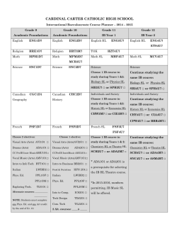
Technical Bulletin #424
Extracellular Matrix Proteins Influence Cell Morphology
and Function in Rat Neural Cultures
Amy Goldberger1 and Paula Flaherty2, Rosa Villalba3, Les A. Riblet3, and Ann Cornell-Bell3
1BD Biosciences, Bedford, MA 01730
2Corning Incorporated, Tewksbury, MA 01876
3Viatech Imaging LLC, Ivoryton, CT 06442
Abstract
The interaction between extracellular matrix (ECM)
and neural cells is important in cell differentiation and
morphogenesis of the nervous system. However, conditions
for culture of neuronal cells have not been optimized for
ECM, and poly-lysine often prevails as the substrate of
choice. Quantitative Ca++ fluorimetry was used to screen
ECM proteins singly and in combination to determine culture
conditions resulting in optimal morphology and function of
rat cortical cells. A fluid handling robot was used to ensure
uniformity in cell plating and reagent dispensing. Agonist
responses in cells from single isolations plated on laminin
followed by human fibronectin (LM/HFN) were significantly
enhanced when compared with cells cultured on LM or HFN
alone, as well as on poly-lysine or uncoated tissue culture
plastic. In addition, differences in morphology between
cultures grown on the various substrates were observed.
Pyramidal neurons exhibited long interconnecting processes
with growth cones on LM/HFN. Neurites in LM or HFN
cultures were significantly shorter and cell bodies formed
aggregates on LM. Elongated neurites were characteristic
of cells grown on poly-lysine. Preliminary studies using
time lapse confocal microscopy with a fluorescent Ca++
dye (Fluo3AM) indicated that glutamate receptor function
also varies in cells cultured on different ECM substrates,
correlating with immunological quantitation of the glutamate
receptor (GluR1). Neural cells grown on LM/HFN displayed
improved cell morphology and receptor function. These
studies indicate that ECM is an important component to
consider in obtaining optimal neural cell cultures.
Introduction
HFN, LM, and other ECM proteins play important roles in
cell surface interactions (Yamada, 1991). HFN mediates cell
adhesion, embryonic cell migration and wound healing, while
LM can promote processes as diverse as axonal outgrowth,
maintenance of polarized epithelial cells and metastasis.
Specialized domains of these molecules or peptide recognition
sequences bind specific cell surface receptors, collagens,
proteoglycans, or other ECMs. Adhesion molecules in the
nervous system are involved in axon elongation and mediate
interactions between growth cones and substrates. In addition
to HFN and LM, the glycoproteins N-cadherin, NCAM,
and other members of the immunoglobulin superfamily play
regulatory roles in neuronal function.
Purpose: To optimize cortical cultures by quantifying
interactions of neural cells in culture with ECM proteins.
Fluorimetry on 96-well plates and confocal imaging of coated
coverslips were used to measure ECM effects on:
• cell morphology
• expression of glutamate receptors
• cell adhesion
• activity of glutamate receptors
• development of dendrites and axons
Materials and Methods
ECM coatings: Corning® BioCoat™ Cell Environments
(Corning Life Sciences) for LM, HFN, PDL, LM/HFN on
coated 96-well Tissue Culture Plates Corning Cat. No.
353075 and 22 mm2 coated glass coverslips were used to
analyze neural cultures from neonatal rat cortex.
Cell culture: Neonatal rat cortex were excised from P0-P1
rat pups, the hippocampus was removed and the cortex
was cleaned of all meninges. Cortices were dissociated in a
solution containing 100 mM CaCl2, 50 mM EDTA, 2 mg
Lcysteine, 200 U/ml papain (Worthington) for 40 minutes
at 37°C. Enzyme activity was stopped with 15 mg trypsin
inhibitor, 15 mg bovine serum albumin in 10 ml in complete
media [DMEM/F12 (phenol red-free, Life Technologies),
10% fetal bovine serum (Life Technologies), pen/strep
solution, 20 mM glucose]. Cells were released by trituration
through a 25 ml pipette. Isolated cells were either plated into
the 96-well plates or settled onto ECM coated coverslips. A
Quadra 96 (Tomtec) fluid handling robot was used to create
uniform cultures containing 25-30,000 cells per well in the
96-well plates. Coverslips were plated with densities of 1,000
cells/cm2.
Effects of ECM coatings: Morphology of neural cells grown
on LM, HFN, PDL, or LM/HFN was studied in a 96-well
format using Phase Contrast Interference Microscopy. Cells
grown on ECM-coated coverslips were imaged using a Nikon
PCM2000 confocal scanning laser microscope. Cells were
stained with Fluo3AM and dye was excited at 488 nM with
an Argon laser. Images were printed using an Epson 600
color printer. Immunohistochemistry was used to quantify
the fluorescence levels of labeled neurons and glial cells in
a culture, adhesion to an ECM coating, presence of actincontaining processes as well as dendrites and axons, and
expression levels of the glutamate receptor.
Stained with:
Neurons Neurotag - Boehringer Mannheim
Glial Cells anti-GFAP (glial marker) - Boehringer Mannheim
Adhesion anti-FAK (focal adhesion kinase) - Transduction Laboratories
F-Actin Phalloidin - Molecular Probes
Dendrites Map2 (microtubule associated protein) - Boehringer Mannheim
Axons Tau - Boehringer Mannheim
Cells were fixed (5 minutes each) with 30% EtOH/1% acetic
acid, 60% EtOH/1% acetic acid and 95% EtOH/1% acetic
acid to maintain antigenicity. Cells were permeabilized with
TritonX100 (1% for 1 minute) then incubated with the
primary antibodies overnight at 4°C except for Neurotag
and phalloidin which were incubated for 2 hours and 5
minutes, respectively. A secondary antibody with a direct
fluorescent tag was used to label the bound primary antibody
Fluorescence was quantified using a CytoFluor® II fluorimeter
(Life Technologies). Immunohistochemical staining of these
antibodies was viewed on the Nikon PCM2000 confocal
scanning laser microscope. Ca2+ imaging and fluorimetry was
used to quantitate neurotransmitter receptor activity of cells
grown on different ECM coatings. When receptors are bound
at the cell surface, there is a resulting
Ca2+ spike which can be measured using a Ca2+ fluorescent
dye, Fluo3AM. Cultures were loaded with Fluo3AM (2 uM)
for 40 minutes at 37°C. The 96-well coated plates were rinsed
with saline using the robot and were placed on the fluorimeter
to obtain a “control” reading. Neurotransmitter solutions
(Glutamate, Kainate, GABA, and Acetylcholine, 100 uM) were
then added to the wells and a second “agonist” reading was
taken. Results were expressed as % Change in Fluorescence.
These same Ca2+ transients were viewed in time lapse videos
using the Nikon PCM2000 confocal microscope. At this level of
resolution, individual neurons and glial cells were identified and
localized immunohistochemical labeling was recorded.
ECM Affects Morphology
PDL: Neurons highly branched with very long processes.
Astrocytes show similar process elongation.
LM: Many multipolar and bipolar neurons with processes
which have secondary branches starting on peripheral
dendrites. Astrocytes flattened and spread.
HFN: More types of neurons: multipolar, bipolar, and
pyramidal. Fewer dendrites per cell with branches starting
closer to the cell body. Astrocytes have spread.
LM/HFN: Neurons have thicker dendrites, multiple spines,
secondary branches, and display growth cones. All neuronal
types present. Networks are established. Astrocytes have
spread.
Phase contrast interference micrographs of neural cultures (5 days in vitro) grown on PDL, LM, HFN, and LM/HFN ECM proteins.
Confocal Images on Different ECMs
Confocal Images on Different ECMs Show the Morphology
In greater Detail. Neurons on Pdl Coating Show very Long
Processes With Extensive Branching (Top). Lm Coating
Shows Neurons With long Processes With few Secondary
Branches (Bottom).
Confocal Images on Different ECMs show Long processes
On neurons Grown on hfn (Top). Dendritic Branching Occurs
close to Cell body. Neurons grown On lm/hfn Show very
long Extensive Processes with Secondary Branches (Bottom).
Quantification of Neurons
Neurotag Staining
350
Fluorescence Units
(+/- S.E., n=6 wells)
300
250
200
150
100
50
0
Laminin
Human Fibronectin
ECM
Poly-D-Lysine
Laminin/Human
Fibronectin
ECM does not affect neuron profile. Similar Neurotag staining on all coatings was measured using fluorimetry (left). Confocal micrograph shows
characteristic dendritic branches close to the cell body when neurons are grown on HFN (right).
Quantification of Glial Cells
GFAP Staining
Fluorescence Units
(+/- S.E., n=6 wells)
1250
1000
750
500
250
0
Laminin
Human
Fibronectin
Poly-D-Lysine
ECM
Laminin/Human
Fibronectin
ECM does not affect glial cell profile. All coatings had similar glial profiles as judged by GFAP fluorescence (left). Confocal immunohistochemistry shows
the complex morphology of astrocytes grown on HFN (right).
Quantification of Adhesion
FAK Antibody Staining
Fluorescence Units
(+/- S.E., n=12 wells)
1000
750
500
250
0
Laminin
Human
Fibronectin
Poly-D-Lysine
ECM
Laminin/Human
Fibronectin
ECM affects cell adhesion. Anti-Focal Adhesion Kinase antibodies
localized adhesion plaques in 96-well plates coated with different
ECMs. Fluorimetry revealed that coatings containing LM and HFN had
greater fluorescence due to FAK (left). PDL was less adhesive. FAK
was localized to large plaques in neuronal cell bodies (middle) and to
plaques throughout the astrocyte cytoplasm (right).
Quantification of Dendrites
MAP-2 Antibody Staining
800
Fluorescence Units
(+/- S.E., n=6 wells)
700
600
500
400
300
200
100
0
Laminin
Human Fibronectin
ECM
Poly-D-Lysine
Laminin/Human
Fibronectin
ECM affects dendrite outgrowth. Anti-map2 higher on HFN coatings or combinations shown by fluorimetry (left). There is significantly less MAP2
fluorescence on PDL coatings. Confocal images show MAP2 along dendrites and in neuronal cell bodies (right).
Quantification of Axons
Tau Antibody Staining
800
Fluorescence Units
(+/- S.E., n=6 wells)
700
600
500
400
300
200
100
0
Laminin
Human Fibronectin
Poly-D-Lysine
ECM
Laminin/Human
Fibronectin
ECM affects axon outgrowth. Anti-tau staining lower on pdl coating seen with fluorimetry (left). There are significantly fewer axons on pdl. Confocal
images show positive tau staining in very thin axons and in nerve cell bodies (right).
Quantification of Actin
Phalloidin Staining
350
Fluorescence Units
(+/- S.E., n=6 wells)
300
250
200
150
100
50
0
Laminin
Human Fibronectin
Poly-D-Lysine
ECM
Laminin/Human
Fibronectin
PDL influences actin polymerization. Phalloidin binding was used to quantitate f-actin in cells grown on ECMs (left). Cells grown on pdl coating exhibited
significantly higher fluorescence from f-actin staining quantified with fluorimetry. Confocal images show the filopodia on neurons and lamelliopodial
veils on astrocytes (right). Morphology is shown for PDL coating, Fluo3AM dye.
1800
Quantification of Glutamate Receptor
GluR1 Antibody Staining
Fluorescence Units
(+/- S.E., n=12 wells)
1600
1400
1200
1000
800
600
400
200
0
Laminin
Human Fibronectin
ECM
Poly-D-Lysine
Laminin/Human
Fibronectin
ECM affects glutamate receptor expression. Lower glutamate receptor expression on pdl was judged by Fluorimetry using anti-glur1 receptor antibodies
(left). High levels of glutamate receptor seen on cells grown on Lm/hfn (right). Staining extends into small neuronal processes.
Quantification of Immunofluorescence
Fluorescence units are expressed as Mean +/- S.E. for readings of 8 wells.
ECM Coating
Stain/Fluorescence
Anti-GFAP
LM/HFN S.E. HFN S.E. LM S.E. PDL S.E.
1012.67
46.37
1038.33
40.76
937
32.96
1024.3 26
269
17.03
293
13.65
297.16
7.37
291.17 10.14
825
31.3
837.84
22.5
826.67
36.4
658
32.8
773.33
22.3
767.67
21.6
705
20.77
594
29.57
740
23.32
739
14.12
693
30.08
577.2
11.8
217.6
3.74
243.5
13.85
239.83
3
299.83 32
1665.33
32.24
1669.68
37.97
1598.33
37.13
1349
(Glial Cells)
Neurotag
(Neurons)
Anti-FAK
(Focal Adhesion Kinase)
Anti-MAP2
(Dendrites)
Anti-Tau
(Axons)
Phalloidin
(F-Actin)
Anti-GluR1
(Glutamate Receptor)
Summary of Immunofluorescence Results:
• Cell numbers do not vary significantly on the different ECMs
• FAK staining is lowest on cells grown on PDL
• MAP2 staining is highest on LM/HFN and on HFN and lowest on PDL
• Tau staining is highest on LM/HFN and on HFN and lowest on PDL
• F-Actin staining was highest on PDL
• GluR1 staining is highest on LM/HFN and on HFN and lowest on PDL
29.4
% Change in Fluorescence
(+/- S.E., n=30 wells)
25
20
15
10
5
0
40
% Change in Fluorescence
(+/- S.E., n=30 wells)
30
Tissue Culture
Plastic
Laminin
Human
Fibronectin
Poly-D-Lysine
ECM
Glutamate Receptor Response on
Different ECM Coatings
30
25
20
15
10
5
0
Tissue Culture
Plastic
Laminin
Human
Fibronectin
ECM
Poly-D-Lysine
20
15
10
5
40
35
Laminin/Human
Fibronectin
GABA Receptor Response on
Different ECM Coatings
25
0
Laminin/Human
Fibronectin
% Change in Fluorescence
(+/- S.E., n=30 wells)
% Change in Fluorescence
(+/- S.E., n=30 wells)
30
Acetylcholine Receptor Response on
Different ECM Coatings
Tissue Culture
Plastic
Laminin
Human
Fibronectin
Poly-D-Lysine
ECM
Laminin/Human
Fibronectin
Kainate Receptor Response on
Different ECM Coatings
35
30
25
20
15
10
5
0
Tissue Culture
Plastic
Laminin
Human
Fibronectin
ECM
Poly-D-Lysine
Laminin/Human
Fibronectin
ECM affects neurotransmitter receptor function. Fluo3AM fluorescence was used to quantitate neurotransmitter receptor activity of neural cells on
different ECMs. Robot-made cultures were used to establish uniform cultures (read day 4 in vitro). The greatest responses to glutamate acetylcholine and
gaba were observed on lm/hfn (left).
Glutamate receptor function on LM/HFN. Time-lapse confocal videos show Ca2+ levels increase with activity of the glutamate receptor. Left Panel: saline,
Right Panel: 100 μM Glutamate. Ca2+ transients increase in neuronal processes as well as cell bodies (Fluorescence=blue<green<yellow<orange).
Conclusion
1. ECMs affect process outgrowth, branching of neurites, and profile of neural cells. LM and LM/HFN coatings support cells with the most diverse morphologies.
2.
ECMs affect adhesion, LM and HFN coatings or combinations have greater adhesion.
3. ECMs affect expression and growth of axons and dendrites. Greatest number of dendrites and axons are seen on HFN and LM/HFN coatings.
4. ECMs affect polymerization of actin filaments. High levels of F-Actin are produced on PDL without concomitant increase in the number of dendrites or axons.
5. ECMs affect expression of the glutamate neurotransmitter receptor. The highest levels of GluR1 are found on LM, HFN, and LM/HFN.
6. ECMs directly affect neurotransmitter receptor activity and function. For glutamate, the greatest activity was seen on LM/
HFN. For kainate, cells grown on HFN and LM/HFN have the highest activity levels. For acetylcholine and ABA, cells grown on LM/HFN have substantially higher activity levels.
Corning acquired the BioCoat™ brand.
For information, visit www.corning.com/discoverylabware.
For Research Use Only. Not for use in diagnostic or therapeutic procedures.
For a listing of trademarks, visit us at www.corning.com/lifesciences/trademarks.
All other trademarks are property of their respective owners.
Corning Incorporated, One Riverfront Plaza, Corning, NY 14831-0001
Corning Incorporated
Life Sciences
836 North St.
Building 300, Suite 3401
Tewksbury, MA 01876
t 800.492.1110
t 978.442.2200
f 978.442.2476
www.corning.com/lifesciences
© 2012, 2013 Corning Incorporated Printed in USA 3/13 CLS-DL-CC-051
ECM coatings profoundly affect neural cell morphology as well as function.
© Copyright 2026 ExpyDoc