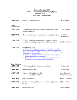
Palaeodiversity 8: 89–94; Stuttgart 30 December 2015.
89
Two new species of Aeolothripidae from Baltic Tertiary
amber (Insecta: Thysanoptera)
MANFRED R. ULITZKA
Abstract
The present study deals with two fossil thrips species of the family Aeolothripidae (Insecta: Thysanoptera),
Rhipidothripoides juttae sp. n. and Mymarothrips groehni sp. n. Both are preserved as inclusions in Baltic Tertiary
Amber (Eocene). Furthermore, a method to facilitate microscopic examinations by embedding the amber in synthetic resin is suggested.
K e y w o r d s : Fossil Thysanoptera, Aeolothripidae, Mymarothrips, Rhipidothripoides, Baltic Tertiary amber,
conservation of amber.
1. Introduction
The Thysanoptera family Aeolothripidae contains
about 200 extant species placed in 23 genera (THRIPSWIKI
2015). Considering the present diversity the number of fossil Aeolothripids appears rather rare: up to now 13 species
(7 genera) have been described from different geological
eras. These fossils range from the Permian (Permothrips
longipennis MARTYNOV, 1935 [SHAROV 1972]) and Cretaceous (Fusithrips crassipes SHMAKOV, 2009 and Cretothrips antiquus GRIMALDI et al., 2004) to the Tertiary.
Most of the fossils associated with Aeolothripidae have
been originated from Tertiary layers where they have
been found mainly as imprints in lime, potash and lignite
(PRIESNER & QUIEVREUX 1936; VON SCHLECHTENDAHL 1887;
SCUDDER 1875; SHMAKOV 2014). Due to their poor preservation in these matrices, their attribution on family status
remains in large parts doubtful and requires review. From
Baltic Tertiary (Eocene) amber, which presents a wide
range of insect inclusions in excellent condition, only one
single genus is known: Rhipidothripoides BAGNALL, 1923.
At present, this genus includes two species described by
monotypy of single female specimens (BAGNALL 1923;
SCHLIEPHAKE 2001).
More than 50% of the extant Aeolothripidae genera are
restricted to tropical countries (MOUND et al. 2012). Due to
the warm and very humid climates in the bygone amber
forests (GRÖHN 2013) these insects may have found similar
living conditions just as in today’s tropics. It is therefore
realistic to believe that Aeolothripids were widespread in
these ecosystems. Furthermore, it might be assumed that
they had shown similar behavioural patterns as their related
extant descendants, which appear to be obligate predators
either high in the trees’ canopy or at ground level.
The two species described below belong to the genera Mymarothrips BAGNALL, 1928 and Rhipidothripoides
BAGNALL, 1923. Rhipidothripoides is an exclusively fossil
genus that was set up by BAGNALL (1923) in conformity
of antennal structures to extant Rhipidothrips UZEL, 1895.
Having antennal segments VII–IX connate, but the forewing with well developed cross veins and both longitudinal veins joining the ring vein well before the wing apex,
it occupies a position between Melanthrips HALIDAY, 1836
and Rhipidothrips. (MOUND 1968; MOUND & MARULLO
1998; ZUR STRASSEN 2003). Mymarothrips in contrast up
to now contains only extant species spread over the Old
World tropics of Africa, India, China, Indonesia, and Australia. Concerning its morphologic structures, the fossil
specimen corresponds considerably with the extant members of that genus. The invention of a possibly new fossil
genus appeared useless therefore. According to MOUND &
MARULLO (1998) the classification within extant Mymarothrips is not yet clear. Three species are currently listed
but these possibly could represent local variants of one
single species that occur with a range of intraspecific variation. However, it is not the purpose of the present paper
to clarify taxonomic obscurities within that genus. The
described specimens ipso facto appear interesting, revealing a first male of Rhipidothripoides and a first fossil of
Mymarothrips.
Acknowledgements
I wish to thank CARSTEN GRÖHN for many fruitful discussions about amber inclusions and especially for ceding me the
specimen of M. groehni for scientific analysis. I thank ANDREA
HASTENPFLUG-VESMANSIS (Senckenbergische Naturforschende
Gesellschaft, Frankfurt am Main, Germany) for sending microscope slides of extant species of Mymarothrips. I want to express
further sincere thanks to CLAIRE MELLISH and the photographer
HARRY TAYLOR (both Natural History Museum, London, U.K.)
for a virtual loan of Rhipidothripoides abdominalis with very
detailed photos. For the fabrication of precise silicone moulds
that facilitate and accelerate the process of making the XOR
90
PALAEODIVERSITY 8, 2015
Crystal-Resin casts a lot, I would like to thank my colleague
ANDREAS K LAPKO. I thank my colleague K IRSTEN GORA for proofreading the manuscript. For the detailed review of this paper
I am very thankful to Prof. Dr. GERALD B. MORITZ (MartinLuther-University Halle-Wittenberg, Germany) and Dr. ANDRÉ
NEL (Muséum National d’Histoire Naturelle, Paris, France).
2. Material and methods
The present study deals with two amber inclusions of the
Thysanoptera collection of the author. Originally these fossils
derived from areas around the Baltic Sea. The inclusion of the
Mymarothrips was generously ceded to the author by CARSTEN
GRÖHN (formerly Coll. GRÖHN No. 3770 W, now: Coll. ULITZKA
No. MU-Fos 36/1), the Rhipidothripoides fossil (Coll. ULITZKA
No. MU-Fos 21/1) was bought in December 2013 from MARIUS
VETA (Palaga, Lithuania). Both specimens are deposited in
the Thysanoptera collection of the Senckenbergische Naturforschende Gesellschaft in Frankfurt am Main, Germany (reference numbers below).
To prepare the fossils for the microscopic examination the
amber first was cut and polished to slices of about 3 mm thickness. Centring the thrips inclusions midway, these slices were
embedded in XOR-Crystal-Resin and finally ground again and
polished with a water-fed flat lap to the size of a standard microscope slide (76 x 26mm) and a thickness of at most 2 mm (Fig.
1). As not only the synthetic resin but also the amber was wet
again by this procedure, the complete preparation was varnished
with Acrüdur R 40 single-component polyurethane resin (Adolff
C.C. Rüegg GmbH & Co Hamburg, Germany). This method of
embedding the amber in such thin microscope slide-like arti-
ficial resin blocks permits accurate front and rear view of the
bioinclusion (see Figs. 2 and 3). The resin stabilizes the amber
and prevents oxidation. Furthermore, centring the tiny inclusions makes its handling much more suitable during the microscopic examinations, just as the comfortable size of standard
microscope slides, which over and above simplifies storing these
samples in normal slide boxes (see also ULITZKA 2015).
All examinations were carried out using a Zeiss standard microscope with the following objectives: Zeiss Plan 10/0,
22 160/-, Nikon M Plan 20x ELWD 210 mm and Nikon M Plan
40x ELWD 0.5NA 210 mm. Illumination was implemented
merging transmission light with two or three white-light-LED
incident illuminators. White paper was used as a diffusor for
incident illumination to prevent reflections in the amber; different coloured paper sheets were inserted under the slide with
transmission light to get the inclusion in better contrast to the
yellowish translucent amber. Photo micrography was performed
with a digital camera attached to the microscope (Canon EOS
70d). The presented photos were produced in focus stacking
technique with Helicon Focus software. Nik Sharpener Pro and
Adobe Photoshop CS3 were used for final colour adjustment and
sharpening. Subsequently, details of the thrips inclusions were
drawn using a Zeiss drawing tube attached to the microscope.
3. Descriptions of the new species
Rhipidothripoides juttae sp. n.
Figs. 2, 3, 7, 8
E t y m o l o g y : This gracile species named “juttae” is dedicated in love and as a wedding gift to my fiancée and partner for
Fig. 1. Microscope slide-like block of polished XOR-Chrystal-Resin containing the midway centred amber inclusion of Rhipidothripoides juttae sp. n. and Mymarothrips groehni sp. n. Measurements in mm (length/width/thickness): 76 x 26 x 1.8.
ULITZKA: NEW SPECIES OF AEOLOTHRIPIDAE FROM BALTIC AMBER
Fig. 2. Rhipidothripoides juttae sp. n. ♂ (ventral view) close to
syninclusions of an aphid and a stellate hair presumably of an
oak tree.
Fig. 3. Rhipidothripoides juttae sp. n. ♂.
many years, JUTTA MOROZ. She encouraged me countless times
to keep on studying thrips species and supports all my scientific
work on these insects.
M a t e r i a l s t u d i e d : Holotype male; Lithuania, Baltic
Sea, bought from MARIUS VETA (Palanga, Lithuania); Collection
ULITZKA No. MU-Fos 27/1; deposited in the Senckenbergische
Naturforschende Gesellschaft: SMF T 19262).
G e n d e r : Male.
D e s c r i p t i o n : Colour difficult to assess. At least head,
prothorax and distal abdominal segments appear to have been
dark brown (Fig. 3); remaining parts of the body and legs light
brown; basal segments of all tarsi distally with a narrow ringlike dark brown shading. Antennae (Fig. 7) light brown; segments III–VII distally dark brown; segments VIII and IX dark
brown. Wings without any visible shading; fringes of forewing
dark brown.
Head (Fig. 8) slightly retracted under pronotum; wider than
long, transversely sculptured on posterior half and produced in
91
front of the eyes; antennae attached more ventrally, therefore
antennal segment I and base of II not visible from above; with
2 pairs of small ocellar setae and 3–4 pairs of minute postocular setae close to the inner hind margin of the compound eyes;
vertex without any setae. Compound eyes large and protruding
in front of convex cheeks; fore ocellus protruding and directed
forwards. Mouth cone short and pointed. Maxillary palps 3-segmented; labial palps 4-segmented. Antennae (Fig. 7) about half
as long as body; 9-segmented; segment II cup-shaped, segments
III–VI parallel-sided and elongate, segment VII elongate coneshaped and connate to segments VIII and IX that are forming a
short 2-segmented conical style; segments III–VII bearing many
prominent setae that are longer than the diameter of the antennal
segments; segment IV with a small oval sensorium on apex (sensoria on segments III not visible); each of the segments V and VI
bearing one slender sense cone. Prothorax (Fig. 8) wider than
long. Pronotum smooth; without any longer setae. Mesonotum
with 2 pairs of minute setae on its posterior margin. Metanotum as far as visible with faintly elongate sculpture. Wings
(Fig. 3) with microtrichia; distally broadly rounded; forewings
with 4 cross veins; costa with about 27 minute setae, anterior
vein (radius) bearing a row of 14 small setae and posterior vein
(media) bearing a row of 12 setae; both longitudinal veins join the
ring vein well before the wing apex; posterior fringes straight.
Legs long and slender; all femora and tibiae with rings of microtrichia; all basal tarsal segments about three times as long as
broad; hamus on the fore tarsi not visible due to the position
of the legs. Abdomen distally with long delicate setae; abdominal tergite I with two diverging longitudinal ridges; microtrichia visible laterally on tergites II–VII; tergite VIII completely
covered with rows of dense microtrichia; hind edge of tergite IX
medially with a comb-like structure bearing tiny teeth.
M e a s u r e m e n t s (holotype male, in microns): Body
length 705 (slightly contracted). Head, length 105; width across
the eyes 140; width behind the eyes 124. Eyes, length 50, width
47. Hind ocelli length 19, width 14; distance between the hind
ocelli 19. Ocellar setae about 8; postocular setae about 11. Pronotum, length 101; middle width 136; pterothorax, largest width
213. Abdomen, length 388; largest width (at base of segment III)
144; abdominal tergite I, length 74; setae on segments IX and
X 43–58. Antennae, length 352; length (width) of segment I 16
(27), II 39 (23), III 66 (16), IV 70 (16), V 58 (16), VI 50 (16), VII
39 (basally 14, distally 10), VIII 8 (5), IX 6 (4). Forewings, length
600; width in the middle 43. Forelegs, length of femora 132, tibiae 124, basal segments of tarsi 35; mid legs, length of femora
132, tibiae 140, basal segments of tarsi 35; hind legs, length of
femora 160, tibiae 167, basal segments of tarsi 50.
D i f f e r e n t i a l d i a g n o s i s : Members of the genus
Rhipidothripoides have antennal segments VII–IX connate
as in Rhipidothrips but the forewing has well developed cross
veins and both longitudinal veins join the ring vein well before
the wing apex as in Melanthrips (MOUND, 1968). Presenting
this conspicuous combination of characteristics, the new species is clearly attributable with Rhipidothripoides. Up to now,
two species have been described in that genus: R. abdominalis
BAGNALL, 1923 and R. involvus SCHLIEPHAKE, 2001, each one
based on a single holotype female. Further specimens are not
known or remained unpublished. Whereas in R. abdominalis
the ocellar setae as well as all prothoracic setae are well developed, these setae are minute or lacking in R. involvus (BAGNALL
1923; MOUND 1968; SCHLIEPHAKE 2001). The new species resembles R. involvus in the missing of the major setae; however, its
antennae are conspicuously longer even though its body length
92
PALAEODIVERSITY 8, 2015
is much smaller. Furthermore, in R. involvus the antennal segments III–VI are not completely parallel sided but slightly convex. Even assuming gender-dependent varying size ratios linked
to an intraspecific sexual dimorphism concerning the antennae,
R. involvus differs in having the compound eyes much larger,
occupying nearly the complete sides of the head. Besides this, in
R. juttae sp. n. the head shape is completely different: the head is
constricted behind and produced in front of the compound eyes;
the fore ocellus is protruding and directed forwards. These characteristics and the visual morphological appearance of the holotype female of R. involvus (as far as assessable due to the milky
covering of this specimen) refute the presumption that the present specimen could represent an according male.
S y n i n c l u s i o n s : One non-identified aphid (Insecta:
Aphidoidea) and some stellate hairs presumably from oak trees
(Fig. 2).
Fig. 4. Mymarothrips groehni sp. n. ♂.
Mymarothrips groehni sp. n.
Figs. 4–6
E t y m o l o g y : This remarkable species is named
“groehni” to honour the research work on amber of CARSTEN
GRÖHN (first chairman of the Verein zur Förderung des
Geologisch-Paläontologischen Museums der Universität Hamburg e.V., “Arbeitskreis Bernstein”). He was the owner of the
specimen before description.
M a t e r i a l s t u d i e d : M. groehni sp. n. holotype female;
Baltic Sea, exact origin not known; Collection ULITZKA No.
MU-Fos 36/1; deposited in the Senckenbergische Naturforschende Gesellschaft: SMF T 19263).
G e n d e r : The sex of this specimen is at first difficult to
assess. The assumption that it is a female is supported by the
intraspecific sexual dimorphism of extant Mymarothrips species: They have sensoria on antennal segments III and IV transverse around the apex ventrally in females, but prolonged
dorso-laterally to near the base of the segment and then recurved
ventrally in males (BHATTI 2006; MOUND & MARULLO 1998). M.
groehni corresponds in these structures with extant females
and therefore its sex is clearly decidable. The barely perceptible
pointed structure could also be interpreted as the distal part of
an ovipositor that is retracted in the abdomen.
D e s c r i p t i o n : Colour of body and head light brown
(Fig. 4), colour of the antennae not easy to assess: light brown,
segments VII–IX on the left antenna appear slightly darker. All
major setae pale. Forewings with traces of a presumably broad
median shading (not clearly assessable); fringes pale.
Head wider than long; not produced in front of the eyes.
Ocelli and eyes large; compound eyes not distended ventrally.
Ocellar setae removed, but at least the points of insertion of
two pairs visible; vertex with three pairs of very small postocular setae. Mouth cone short. Maxillary palps 3-segmented.
Antennae (Fig. 5) about one third as long as body, 9-segmented;
segments III–VII moniliform, but almost rectangular, with
prominent setae and slender pedicels at base; VIII cone-shaped;
IX slender, forming a slightly cone shaped pedicle. Segments III
and IV each with a ventral transverse kidney-shaped sensorium
around the apex. Prothorax much wider than long. Pronotum
smooth except for some faint lines of transverse sculpture close
to the lateral margins; without longer setae except for one pair
of prominent posteroangular setae. Pterothorax not clearly visible; at least metanotum transversely sculptured. Forewings typi-
cally Mymarothrips-shaped (Fig. 6), with base slender, but apex
broadly rounded; covered with microtrichia; forewings with
4 cross veins; costa with 29 setae; anterior vein (radius) bearing 8 setae between its base and the median cross vein (distal
setae not visible), posterior vein (media) bearing about 13 setae.
Fringes of the forewing short and straight. Legs long and slender; tarsi two segmented. Basal segments of abdomen not visible
(covered by wings), distal segments without microtrichia; sternites without discal setae.
M e a s u r e m e n t s (holotype female, in microns):
Body length 858 (slightly contracted). Head, length about 150
(retracted under pronotum, therefore difficult to measure); width
across the eyes 169. Eyes, length 90, width 68. Hind ocelli length
19, width 14; distance between the hind ocelli 15. Pronotum,
length 83; width 165; Abdomen, length 450. Antennae, length
segment III–IX 287 (segments I and II not measurable); length
(width) of segment III 58 (23), IV 49 (20), V 54 (19), VI 45 (19),
VII 47 (20), VIII 17 (basally 16, distally 8), IX 12 (4). Forewings,
length 600; largest width 176. Forelegs, length of femora 135,
tibiae 135, basal segments of tarsi 45; mid legs, length of femora
132, tibiae 135, basal segments of tarsi 60; hind legs, length of
femora 180, tibiae 143, basal segments of tarsi 60.
D i f f e r e n t i a l d i a g n o s i s : As mentioned above, the
genus Mymarothrips currently contains three extant species.
The female of M. groehni sp. n. is the first fossil representative.
Due to its distinctive racket-shaped forewings with the distal
part about twice as broad as the base and due to its characteristic antenna with segments III–VII similar in size and shape, it
can be clearly classified as a member of that genus. Species vary
in the course of time and therefore each of them has a limited
period of existence only (ZUR STRASSEN 1973). Based on this fact
alone the fossil Mymarothrips of the Tertiary era is not attributable to any Recent species. Furthermore, and contrary to all the
extant species, M. groehni sp. n. differs in having only one pair
of pronotal prominent setae (setae posteroangulares). Beside
this, antennal segment VIII is much wider at its base and therefore more cone-shaped than in extant species.
S y n i n c l u s i o n s : A fossil wasp of the family Diapriidae
(Insecta: Hymenoptera), a mite (Arachnida: Acari) and some
stellate hairs presumably from oak trees.
ULITZKA: NEW SPECIES OF AEOLOTHRIPIDAE FROM BALTIC AMBER
93
Figs. 5, 6. Mymarothrips groehni sp. n. ♂. 5: Right antenna, segments III–IX. 6: Right forewing (microtrichia are not shown).
Figs. 7, 8. Rhipidothripoides juttae n. sp. ♂. 7: Right antenna. 8: Head and prothorax. The scale shown above is equal to 250 μm in
Figs. 6 and 8; in Figs. 5 and 7 it is equal to 125 μm.
4. References
BAGNALL, R. (1923): Fossil Thysanoptera I, Terebrantia Pt. I. –
Entomologist’s monthly Magazine, 59: 35–38.
BAGNALL, R. (1928): Preliminary description of Mymarothrips
ritchianus, a new type of Thysanopteron. – Annals and
Magazine of Natural History, 10 (1): 304–307.
BHATTI, J. S. (2006): The classification of Terebrantia (Insecta)
into families. – Oriental Insects, 40: 339–375.
GRIMALDI, D., SHMAKOV, A. & FRASER, N. (2004): Mesozoic thrips
and early evolution of the Order Thysanoptera (Insecta). –
Journal of Paleontology, 78 (5): 941–952.
GRÖHN, C. (2013): Alles über Bernstein. 207 pp.; Neumünster
(Wachholtz Verlag).
MARTYNOV, A. V. (1935): A find of Thysanoptera in the Permian
deposits. – Comptes Rendus (Doklady) de l’Académie des
Sciences de l’URSS, 3 (7): 333–336.
MOUND, L. A. (1968): A review of R.S. BAGNALL’s Thysanoptera
collections. – Bulletin of the British Museum (Natural History), Entomology, 11: 1–181.
MOUND, L. A. & MARULLO, R. (1998): Biology and identification of Aeolothripidae (Thysanoptera) in Australia. – Invertebrate Taxonomy, 12 (6): 92–950.
MOUND, L. A., TREE, D. C. & PARIS, D. (2015): OzThrips – Thysanoptera in Australia. –http://www.ozthrips.org/terebrantia/
aeolothripidae/ [accessed March 2015].
PRIESNER, H. & QUIEVREUX, F. (1936): Thysanopteres des couches de potasse du Haut-Rhin. – Bulletin de la Société Géologique de France, 5 (193): 471–477.
SCHLECHTENDAHL, D. VON (1887): Physopoden aus dem Braunkohlengebirge von Rott am Siebengebirge. – Zeitschrift für
Naturwissenschaften, 60: 551–592.
SCHLIEPHAKE, G. (2001): Thysanoptera (Insecta) of the Tertiary
amber of the Museum of the Earth, Warsaw, with keys to the
94
PALAEODIVERSITY 8, 2015
species of the Baltic and Bitterfeld amber. – Prace Muzeum
Ziemi, 46: 17–39.
SCUDDER, S.H. (1875): The Tertiary Physopoda of Colorado. –
Bulletin of the U.S. Geological and Geographical Survey of
the Territories, 1 (4): 221–223.
SHAROV, A. G. (1972): On the phylogenetic relations of the order
Thripidae (Thysanoptera). – Entomological Review, 54:
854–858.
SHMAKOV, A. S. (2009): The oldest members of the families
Aeolothripidae and Thripidae (Insecta: Thysanoptera) from
the Lower Cretaceous of Transbaikalia. – Paleontological
Journal, 43 (4): 428–432.
SHMAKOV, A. S. (2014): Thrips (Insecta: Thysanoptera) from the
Insect Limestone (Bembridge Marls, Late Eocene) of the Isle
of Wight, UK. – Earth and Environmental Science Transactions of the Royal Society of Edinburgh, 104: 317–325.
ZUR STRASSEN, R. (1973): Fossile Fransenflügler aus mesozoischem Bernstein des Libanon (Insecta: Thysanoptera). –
Stuttgarter Beiträge zur Naturkunde, 256: 1-51.
ZUR STRASSEN, R. (2003): Die terebranten Thysanopteren Europas
und des Mittelmeer-Gebietes. – In: DAHL, F. (Ed.): Die Tierwelt Deutschlands, 74: 277 pp.; Keltern (Goecke & Evers).
ThripsWiki (2015): ThripsWiki – providing information on
the World’s thrips. –http://thrips.info/wiki/Aeolothripidae
[accessed March 2015].
ULITZKA, M. R. (2015): Thrips-iD – Die Thysanopterenseite von
Dr. MANFRED ULITZKA. – http://www.thrips-id.de/Thrips-iDDeutsch/thrips-id-fossil.html [accessed March 2015].
Address of the author:
Dr. MANFRED R. ULITZKA, Zeller Straße 14, D-77654 Offenburg, Germany.
Email: [email protected]. Internet: www.thrips-id.de
Manuscript received: 13 July 2015, revised version accepted: 14 September 2015.
© Copyright 2026 ExpyDoc