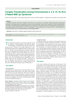
Korean J Crit Care Med
2014 February 29(1):43-47 / http://dx.doi.org/10.4266/kjccm.2014.29.1.43
pISSN: 1229-4802ㆍeISSN: 2234-3261
■ Case Report ■
Delayed Hemolytic Uremic Syndrome Presenting as Diffuse Alveolar Hemorrhage
Ji Young Hong, M.D., Ji Ye Jung, M.D., Young Ae Kang, M.D., Yoon Sung Bae, M.D., Young Sam Kim, M.D.,
Se Kyu Kim, M.D., Joon Chang, M.D., and Moo Suk Park, M.D.
Division of Pulmonology, Department of Internal Medicine, Yonsei University College of Medicine, Seoul, Korea
Hemolytic uremic syndrome (HUS) is defined by the triad of mechanical intravascular hemolytic anemia with schistocytosis, thrombocytopenia and acute renal failure. Pulmonary involvement in HUS is known to be rare. We present the case of a 25-year-old male with
diffuse alveolar hemorrhage and myocarditis followed by atypical hemolytic uremic syndrome. In this case, successful treatments included steroid pulse therapy for the fatal alveolar hemorrhage and plasma exchange for the hemolytic uremic syndrome.
Key Words: hemolytic-uremic syndrome; hemorrhage; plasma exchange.
Diffuse alveolar hemorrhage (DAH) is a life threatening dis-
mmHg. His respiratory rate was 26 breaths/min. There were no
order with high mortality.[1] The treatment of alveolar hemor-
signs of arthritis, skin rashes, or lymphadenopathy. He did not
rhage varies according to the etiologies. The alveolar hemor-
have any abdominal discomfort or diarrhea. Physical examina-
rhage in hemolytic uremic syndrome (HUS) has been rarely re-
tion was unremarkable except for mild crackles over both lower
ported as complication.[2-5] We report a case of atypical HUS
lobes.
which manifested with alveolar hemorrhage, respiratory failure
Initial laboratory findings showed a hemoglobin level of 14.4
and myocarditis even before thrombocytopenia and hemolytic
g/dl, a white blood cell count of 15,000/µl (neutrophil count
anemia developed. We will discuss the causes and treatment of
83.7%), and a platelet count of 134,000/mm3. Chemical values
atypical alveolar hemorrhage in this case review.
were notable for a blood urea nitrogen of 32.2 mg/dl and a creatinine of 2.6 mg/dl (FeNa 7.6%). Urinalysis showed 1+ blood
cells and 2+ proteins. The activated partial thromboplastin time
CASE REPORT
and prothrombin time were in normal ranges. Blood gas analysis
revealed a pH of 7.49, PaCO2 31 mmHg, PaO2 57 mmHg, HCO3-
A 25-year-old man was transferred to the emergency department
with a 4-day history of fever, dyspnea and hemoptysis, despite
23.6 mmol/L, and SaO2 92% while breathing room air. On re-
having received high-dose corticosteroid therapy in a local ter-
verse transcriptase-polymerase chain reaction (RT-PCR) testing
tiary hospital. The patient was a nonsmoker, and did not have
for respiratory viruses, only rhinovirus was found. Immunological
any history of drug use, recent travel, tuberculosis exposure, or a
survey including antinuclear antibody, anti double stranded DNA
significant past medical history.
antibody, antiglomerular basement membrane antibody, anti-
On physical examination, his temperature was 38.0℃, his
neutrophil cytoplasmic antobody were negative. The comple-
heart rate was 116 beats/min, and his blood pressure was 188/89
ment components were normal (C3 130 mg/dl, C4 38 mg/dl).
R.tsutsugamushi antibody, leptospira antibody and Hantaan virus antibody on serologic tests were negative.
Received on January 1, 2014
Revised on January 28, 2014
Accepted on January 28, 2014
Correspondence to: Moo Suk Park, Division of Pulmonology,
Department of Internal Medicine, Yonsei University College of
Medicine, 50 Yonsei-ro, Seodaemun-gu, Seoul 120-752, Korea
Tel: 82-2-2228-1955, Fax: 82-2-393-6884
E-mail: [email protected]
A chest roentgenogram showed bilateral alveolar infiltrates
(Fig. 1a). Chest CT scans showed consolidation and edema with
multifocal ground glass opacities (Fig. 1b). Echocardiography
showed that left ventricular systolic function was reduced to
35%. Severe global hypokinesia of the left ventricle and my43
44 The Korean Journal of Critical Care Medicine: Vol. 29, No. 1, February 2014
A
B
C
D
Fig. 1. (A) Initial chest radiograph. (B) Chest CT scans showed consolidation along the peribronchovascular bundle with
combined multifocal ground glass opacities and interstitial thickening mainly on the central portion of the lung. (C) Gross
findings of bronchoalveolar lavage fluid: the third syringe (right) is obviously more blood-filled compared with the first (left)
syringe. (D) Chest radiograph after steroid pulse therapy.
ocardial edema were observed.
solved (Fig. 1d) and the patient was weaned off. However, the
The patient was placed on IV moxifloxacin following cultures.
renal function deteriorated and the patient underwent a percuta-
On the second day after admission, intubation was performed
neous renal biopsy to distinguish the underlying diseases. The
due to aggravated hypoxic respiratory failure. The steroid pulse
renal biopsy demonstrated that the glomerular capillary loops
therapy was administered under the suspicion of vasculitis in-
showed fibrin thrombi with capillary congestion. On immuno-
volving the kidney, pulmonary alveolar epithelium, and myocardium.
fluorescence, the glomerular sections reacted with antibodies
On fiberoptic bronchoscopy, bronchoalveolar lavage (BAL) flu-
specific for the heavy chains of IgG, IgA, and IgM, and against
id obtained from the lateral basal segment of the right lower lobe
C3, C4, C1q, and fibrinogen (Fig. 2). On the basis of the renal
showed a progressively bloody appearance (Fig. 1c). The bacterial,
biopsy results, he was diagnosed with an atypical hemolytic ure-
fungal and mycobacterial cultures were all negative. Pneumocystis
mic syndrome complicated with alveolar hemorrhage and my-
jiroveci, influenza A, parainfluenza, respiratory syncytial virus,
ocarditis .
cytomegalovirus, and adenovirus were not detected by multi-
On hospital day 7, the platelet count decreased steeply from
plex PCR. Cytologic examination of the BAL fluid was negative
120,000/mm3 to 59,000/mm3, and schistocytes were observed
for malignancy. After the steroid pulse therapy for two days, the
on the peripheral blood smear. Following five days of once daily
alveolar infiltrates on the chest roentgenogram completely re-
plasma exchange treatment, the patient showed a favorable re-
Ji Young Hong, et al: HUS Presenting Diffuse Alveolar Hemorrhage 45
A
B
C
D
Fig. 2. Renal biopsy findings in hemolytic uremic syndrome. (A) Light microscopy showing congestion of glomerular capillary
loops with fibrin thrombi (H & E stain; original magnification × 200). (B) Light microscopy showing renal arteriolar intimal
thickening and narrowing of the lumen (H & E stain; original magnification × 400). (C) Acid fuchsin orange G staining showing
focal fibrinoid deposits and thrombi in the glomerular capillary loops (AFOG × 200). (D) Immunofluorescence showing the glomerular localization of fibrinogen in the mesangium.
sponse, with an increase in hemoglobin, a decrease in microangiopathic hemolysis, improvement of renal function and an
elevation in platelet count to 141,000/µl. The EHEC (Enterohaemorrhagic Escherichia coli) toxin PCR was positive, but a
stool culture was negative. The Positron Emission Tomography
(PET) scan and bone marrow biopsy revealed no evidence of
malignancy or other hematologic diseases. On transthoracic
echocardiography, myocardial edema remained, but left ventricular systolic function improved to 53%. Fig. 3 shows longitudinal changes in the clinical markers according to treatment.
We slowly tapered the steroid dose to 15 mg of oral prednisolone per day after three weeks. After 4 weeks, the patients
was discharged in stable condition and followed up regularly.
DISCUSSION
HUS is classified as either diarrhea-associated/typical HUS
or non-diarrhea/atypical HUS. Typical HUS making up 90 perFig. 3. Time course of hemoglobin, LDH (lactate dehydrogenase), platelet
count, BUN (blood urea nitrogen) and creatinine levels during treatment.
The period of plasma exchange is shown as the gray speckled zone.
cent of all cases, is preceded by gastroenteritis with Shiga-toxin-producing bacteria, ranging from three days to more than two
46 The Korean Journal of Critical Care Medicine: Vol. 29, No. 1, February 2014
weeks.[6] On the contrary, atypical HUS is associated with a va-
and myocarditis could be preceding manifestations prior to the
riety of causes including dysregulation of the alternative com-
development of typical features such as hemolytic anemia or
plement pathway, autoimmune disorders, cancers, drugs (VEGF
thrombocytopenia. It accords with the previous report that al-
inhibitors such as bevacizumab), bone marrow transplantation,
veolar hemorrhage and alveolar wall necrosis can occur even in
and infection.[7] The distinction of pathogenesis is blurred be-
the absence of fibrin thrombus deposition.[12]
cause Shiga toxin can also mediate alternative complement
In atypical HUS, plasmapheresis with infusions of fresh fro-
pathway activation and acquired complement dysfunction in
zen plasma is the first treatment option.[13] Fortunately, our pa-
typical HUS.[8]
tient showed good response to fifth session of plasma exchange.
In this case, no clinical symptoms of typical HUS were pres-
The complement blocker, eculizumab (humanized monoclonal
ent except for the presence of EHEC toxin on PCR. In addition,
anti-C5 immunoglobulin G) should be considered for HUS pa-
the excellent response to plasma exchange raises the possibility
tients who are resistant to plasma exchange.
of atypical HUS. Among the etiologies of atypical HUS, human
In summary, this case highlights uncommon multivisceral
immunodeficiency virus infection, malignancy, and immune-re-
complication of atypical HUS. Pulmonary hemorrhage asso-
lated diseases were excluded. The C3 and C4 levels were nor-
ciated HUS should be considered as an uncommon cause.
mal, but a complete genetic analysis for factor H, factor I, factor
Several investigations including infectious etiology or com-
B, membrane cofactor protein (CD46) and ADMAMTS-13
bined complement dysfunction are required. Kidney biopsy
could not be performed due to lack of utility. It is unclear wheth-
could contribute to distinguish the underlying atypical HUS
er Rhinovirus is an infectious trigger leading to atypical HUS
from other collagen vascular diseases and to start appropriate
along with impaired immune system. Several cases have shown
treatment. Steroid therapy and plasma exchange should be per-
that viruses may be a precipitating trigger in the pathogenesis of
formed in fatal hemorrhage and myocarditis in HUS.
atypical HUS.[9] Influenza A causes hemolysis and erythrocyte
fusion with viral neuraminidase, and produces anti-ADAMTS
ACKNOWLEDGMENTS
13 autoantibodies. Vascular endothelium disruption by coxsackievirus and echovirus results in intravascular coagulation. It was
Financial/nonfinancial disclosures: The authors have re-
reported that enterovirus may play a synergistic role in trigger-
ported to respiratory care that no potential conflicts of interest
ing HUS in VTEC (Verotoxin-producing E.coli) - positive HUS.
exist with any companies/organizations whose products or serv-
Further research will investigate the connection between rhino-
ices may be discussed in this article.
virus and endothelial injury leading to thrombotic microangiopathies.
REFERENCES
The differential diagnosis of DAH includes small vessel vasculitis, immune complex-mediated vasculitis, toxic inhalation,
bone marrow transplantation, infection and drug-associated
1) Newsome BR, Morales JE: Diffuse alveolar hemorrhage.
South Med J 2011; 104: 269-74.
disease.[10] The current hypothesis about alveolar hemorrhage
2) Derebail VK, Parikh P, Jennette JC, Kshirsagar AV: A rare
in HUS is that endothelial damage, the landmark of thrombotic
cause of the pulmonary-renal syndrome: a case of atypical
microangiopathy, results in alveolar wall necrosis, loss of capil-
haemolytic-uraemic syndrome complicated by pulmonary
lary integrity and alveolar hemorrhage. The dramatic resolution
haemorrhage. NDT plus 2008; 1: 417-9.
of infiltration after corticosteroid therapy indicates that DAH
3) Piastra M, Ruggiero A, Langer A, Caresta E, Chiaretti A,
may have been associated with destruction of the pulmonary
Pulitano S, et al: Pulmonary hemorrhage complicating a typ-
vascular endothelium.
ical hemolytic-uremic syndrome. Respiration 2004; 71:
Cardiovascular dysfunction in patients with HUS tends to be
537-41.
underdiagnosed. The mechanisms leading to heart failure are
4) Rhee H, Song SH, Lee YJ, Choi HJ, Ahn JH, Seong EY, et al:
due to volume overload after renal failure or myocardial micro-
Pandemic H1N1 influenza A viral infection complicated by
infarctions caused by thrombi in the cardiac circulation.[11]
atypical hemolytic uremic syndrome and diffuse alveolar
Aggressive treatment including plasma exchange should be ini-
hemorrhage. Clin Exp Nephrol 2011; 15: 948-52.
tiated immediately due to poor prognosis.
It is remarkable that renal insufficiency, alveolar hemorrhage,
5) Garnacho Montero J, Marmesat Ríos I, Leal Noval SR,
González Fernández FJ, Goñi Belzunegui MV, Camacho
Ji Young Hong, et al: HUS Presenting Diffuse Alveolar Hemorrhage 47
Laraña P: [The hemolytic-uremic syndrome: a report of a
10) von Ranke FM, Zanetti G, Hochhegger B, Marchiori E:
case which started as a massive pulmonary hemorrhage]. An
Infectious diseases causing diffuse alveolar hemorrhage in
Med Interna 1990; 7: 416-8.
immunocompetent patients: a state-of-the-art review. Lung
6) Loirat C, Saland J, Bitzan M: Management of hemolytic uremic syndrome. Presse Med 2012; 41(3 Pt 2): e115-35.
7) Westra D, Wetzels JF, Volokhina EB, van den Heuvel LP, van
de Kar NC: A new era in the diagnosis and treatment of atypical haemolytic uraemic syndrome. Neth J Med 2012; 70:
121-9.
8) Petruzziello-Pellegrini TN, Marsden PA: Shiga toxin- associated hemolytic uremic syndrome: advances in pathogenesis
and therapeutics. Curr Opin Nephrol Hypertens 2012; 21:
433-40.
9) Lopes da Silva R: Viral-associated thrombotic microangiopathies.
Hematol Oncol Stem Cell Ther 2011; 4: 51-9.
2013; 191: 9-18.
11) Gami AS, Hayman SR, Grande JP, Garovic VD: Incidence
and prognosis of acute heart failure in the thrombotic
microangiopathies. Am J Med 2005; 118: 544-7.
12) Richardson SE, Karmali MA, Becker LE, Smith CR: The
histopathology of the hemolytic uremic syndrome associated
with verocytotoxin-producing Escherichia coli infections.
Hum Pathol 1988; 19: 1102-8.
13) Rosales A, Riedl M, Zimmerhackl LB: Thrombotic microangiopathy: atypical HUS: current diagnostic and therapeutic approaches. Nat Rev Nephrol 2010; 6: 504-6.
© Copyright 2026 ExpyDoc