1020
UNIT 4
Maintenance of the Body
Milliosmols
r-------------------------------~~
Cortex
Na+
H+
K+
300
CIHC03Outer
medulla
Urea
(a)
(b)
600
H2O
(e)
(e)
(e)
Inner
medulla
1200
(b)
Key:
--+= Active transport
(primary or secondary)
--+= Passive transport
(e)
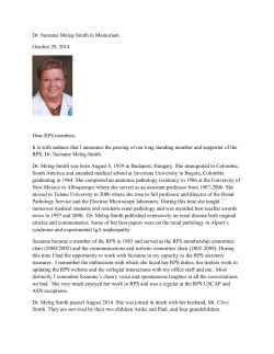
FIGURE 25.16 Summary of
nephron functions. The glomerulus
provides the filtrate processed by the
renal tubule. The various regions of the
renal tubule carry out reabsorption and
secretion and maintain a gradient of
osmolality within the medullary
interstitial fluid. Varying osmolality at
different points in the interstitial fluid
is symbolized in the central figure by
gradients of color. The inserts that
accompany the central figure describe
the main transport functions of the four
regions of the nephron tubule and of
the collecting duct. (a) Proximal tubule.
Filtrate that enters the PCT from the
glomerular capsule has about the same
osmolality as blood plasma. The main
activity of the PCT transport epithelium
is reabsorption of certain solutes from
the filtrate back into the blood. Nearly
all nutrients and about 65% of Na+ are
actively transported out of the PCT and
enter the peritubular capillaries; CI- and
water follow passively. PCT cells also
secrete ammonium and other nitrogenous wastes into the filtrate and help
maintain a constant pH in blood and
interstitial fluid by the secretion of H+
and reabsorption of HC03 -. By the end
(d)
of the proximal tubule, the filtrate
volume has been reduced by 65%.
(b) Descending limb. The descending
limb of the loop of Henle is freely
permeable to water but not to NaCI. As
filtrate in this limb descends into the
medulla, the filtrate loses water by
osmosis to the interstitial fluid, which is
increasingly hypertonic in that direction.
Consequently, salt and other solutes
become more concentrated in the
filtrate. (c) Ascending limb. The ascending limb of the loop of Henle is
impermeable to water but permeable to
Na+ and CI-. These ions, which became
concentrated in the descending limb,
move passively out of the thin portion of
the ascending limb, are actively pumped
out of the thick portion of the ascending limb, and contribute to the high
osmolality of interstitial fluid in the inner
medulla. K+ is cotransported with Na+
and CI-. The tubule epithelium here is
not permeable to water, so the filtrate
becomes more and more dilute as
the exodus of salt from the filtrate
continues. (d) Distal tubule. The DCT,
like the PCT, is specialized for selective
secretion and reabsorption. Na+ and
CI- are cotransported, H+ may be
secreted, and in the presence of
aldosterone, more Na+ is reabsorbed.
The water permeability of the DCT
is extremely low and almost no further H2 0 absorption occurs there.
(e) Collecting duct. The urine, normally
quite dilute at this point, begins its
journey via the collecting duct back into
the medulla, with its increasing
osmolality gradient. In the cortical
collecting duct, K+, W, and/or HC03 ions may be reabsorbed or secreted
depending on what is required to
maintain homeostasis. The wall of the
medullary region of the collecting duct
is permeable to urea and is made more
so by the presence of ADH. Some urea
diffuses out of the collecting duct and
contributes to the high osmolality of the
inner medulla. In the absence of ADH,
the collecting duct is nearly impermeable
to water, and dilute urine is excreted. In
the presence of ADH, more aquaporins
are inserted into the collecting duct, and
the filtrate loses water by osmosis as it
passes through medullary regions of
increasing osmolality. Consequently,
water is conserved, and concentrated
urine is excreted.
Chapter 25
The Urinary System
1021
essentially a sedative, encourages diuresis by inhibiting release of ADH. Other diuretics increase
urine flow by inhibiting Na + reabsorption and the
obligatory water reabsorption that normally follows. Examples include caffeine (found in coffee,
tea, and colas) and many drugs prescribed for hypertension or the edema of congestive heart failure.
Common diuretics inhibit Na+ -associated symporters. ((Loop diuretics" [like furosemide (Lasix)]
are powerful because they inhibit formation of the
medullary gradient by acting at the ascending limb
of Henle's loop. Thiazides are less potent and act at
the DCT.
This is the case with most drug metabolites. Knowing a drug's renal clearance value is essential because
if it is high, the drug dosage must also be high and
administered frequently to maintain a therapeutic
level. Creatinine, which has an RC of 140 mlImin, is
freely filtered but also secreted in small amounts. It
is often used nevertheless to give a ((quick and dirty"
estimate of GFR.
Renal Clearance
Color and Transparency
Freshly voided urine is clear and pale to deep yellow.
Its yellow color is due to urochrome (u'ro-kromJ, a
pigment that results from the body's destruction of
hemoglobin (via bilirubin or bile pigments). The
more concentrated the urine, the deeper the yellow
color. An abnormal color such as pink or brown, or a
smoky tinge, may result from eating certain foods
(beets, rhubarb) or may be due to the presence in the
urine of bile pigments or blood. Additionally, some
commonly prescribed drugs and vitamin supplements alter the color of urine. Cloudy urine may indicate a urinary tract infection.
Renal clearance refers to the volume of plasma that
is cleared of a particular substance in a given time,
usually 1 minute. Renal clearance tests are done to
determine the GFR, which allows us to detect
glomerular damage and follow the progress of renal
disease (discussed in A Closer Look, p. 1022).
The renal clearance rate (RC) of any substance,
in mlImin, is calculated from the equation
RC = UV/P
where
U = concentration of the substance in urine (mglml)
v=
flow rate of urine formation (ml/rnin)
P = concentration of the substance in plasma (mglml)
Because it is freely filtered and neither reabsorbed nor secreted by the kidneys, inulin (in'u-lin)
is the standard used to determine the GFR. A polysaccharide with a molecular weight of approximately
5000, inulin's renal clearance value is equal to the
GFR. When inulin is infused such that its plasma
concentration is 1 mg/ml (P = 1 mglmlJ, then generally U = 125 mg/ml, and V = 1 mlImin. Therefore,
its renal clearance is RC = (125 x 1)/1 = 125 mlImin,
meaning that in 1 minute the kidneys have removed
(cleared) all the inulin present in 125 ml of plasma.
A clearance value less than that of inulin means
that a substance is partially reabsorbed. An example
is urea with an RC of 70 mlImin, meaning that of
the 125 ml of glomerular filtrate formed each
minute, approximately 70 ml is completely cleared
of urea, while the urea in the remaining 55 ml is recovered and returned to the plasma. If the RC is zero
(such as for glucose in healthy individuals J, reabsorption is complete or the substance is not filtered.
If the RC is greater than that of inulin, the tubule
cells are secreting the substance into the filtrate.
Urine
Physical Characteristics
Odor
Fresh urine is slightly aromatic, but if allowed to
stand, it develops an ammonia odor as bacteria metabolize its urea solutes. Some drugs and vegetables
alter the usual odor of urine, as do some diseases.
For example, in uncontrolled diabetes mellitus the
urine smells fruity because of its acetone content.
pH
Urine is usually slightly acidic (around pH 6), but
changes in body metabolism or diet may cause the
pH to vary from about 4.5 to 8.0. A predominantly
acidic diet that contains large amounts of protein
and whole wheat products produces acidic urine. A
vegetarian (alkaline) diet, prolonged vomiting, and
bacterial infection of the urinary tract all cause the
urine to become alkaline.
Specific Gravity
Because urine is water plus solutes, a given volume
has a greater mass than the same volume of distilled
water. The ratio of the mass of a substance to the
mass of an equal volume of distilled water is its
specific gravity. The specific gravity of distilled water is 1.0 and that of urine ranges from 1.001 to
1.035, depending on its solute concentration.
25
1022
UNIT 4
Maintenance of the Body
Chronic Renal Disease: A National Health Crisis in the Making
H
oW long has it been since you had
a urinalysis? If you're like most of
us, the answer is: too long. This
simple, inexpensive, painless test can
detect renal disease years before any
symptoms appear, buying time for
early treatment and preventing lifethreatening complications.
Unfortunately, urinalysis is often
omitted from routine medical checkups. Unfortunate, because chronic renal disease is a major burden to the
U.s. health care system. Some 10-20
million Americans-one out of every
15-have some degree of kidney dysfunction. More than 2.6 million Americans suffer major impairment, and
nearly 350,000 require dialysis or a
kidney transplant just to stay alive.
In addition to its physical toll on
individuals, chronic renal disease
creates a huge financial burden for
the nation. The U.S. spends $22.8 billion annually to treat these patients,
a figure that is projected to increase
to $38.35 billion by 2010. Medicare
spending for renal failure rises by
5-10% every year. People with chronic
kidney disease make up only 0.6% of
the Medicare population, but consume 6% of its budget.
Stages of Renal Disease
25
Renal failure can strike as an acute crisis, perhaps due to trauma, infections,
or poisoning by heavy metals or organic solvents. More often, though, it
develops silently and insidiously over
many years. Filtrate formation decreases gradually, nitrogenous wastes
accumulate in the blood, and blood
pH drifts toward the acidic range.
Chronic renal disease refers to this
gradual loss of kidney function, defined as either kidney damage or a
GFR of less than 60 mllmin for at least
three months. Kidney damage is defined as the presence of structural or
functional abnormalities, or of damage
markers such as abnormal components
in blood and urine.
Clinicians classify chronic renal disease into five stages according to the
level of kidney function:
• Stage 1: Signs of kidney damage
with GFR2:90
• Stage 2: Signs of kidney damage
with G FR of 60-89
• Stage 3: GFR 30-59
• Stage 4: GFR 15-29
• Stage 5: GFR <15
Patients in stage 5 are considered
to be in renal failure (also called endstage renal disease). At this point they
have only 10-15% of kidney function
left, and most require dialysis or a
transplant to survive.
Symptoms and Risk Factors
Warning signs of kidney trouble may
include high blood pressure, frequent
urination, difficult or painful urination,
puffy eyes, or swollen hands or feet.
But all too often, there are few noticeable symptoms until advanced stages,
when a significant percentage of kidney function has already been lost.
Urinalysis is valuable because it can
detect proteinuria, a sensitive early
marker of kidney damage.
The most abundant protein found
in urine is albumin. Normally urinary
Chemical Composition
Water accounts for about 95% of urine volume; the
remaining 5% consists of solutes. The largest component of urine by weight, apart from water, is urea,
which is derived from the normal breakdown of
amino acids. Other nitrogenous wastes in urine include uric acid (an end product of nucleic acid metabolism) and creatinine (a metabolite of creatine
albumin averages less than 100 mg/day
(as measured over a 24-hour period);
excretion of more than 300 mg/day
signals clinical proteinuria.
Other tests that can evaluate kidney
function include
• Creatinine clearance, which measures filtering efficiency by comparing
the creatinine level in the blood to that
in the urine.
• Serum creatinine, which detects
levels of creatinine in the blood.
• Blood urea nitrogen (BUN), which
measures the amount of urea in the
blood. Generally, if creatinine is abnormal, BUN will be too.
Where did this kidney crisis come
from? In adults, chronic renal disease
often develops in conjunction with
other chronic health conditions. As the
U.S. population ages and gains weight
(see A Closer Look in Chapter 24), risk
factors for renal problems multiply.
The leading cause is diabetes mellitus, which accounts for approximately
44% of new cases each year. Hypertension is a close second, accounting for
about 28% of cases. Note that hypertension is both a cause and a symptom: High blood pressure impairs
kidney function by damaging renal
blood vessels and reducing circulation
to the organs, even as hypertensionbattered kidneys push blood pressure
upward. Atherosclerosis compounds
the problem by further impairing circulation.
Race and ethnicity may playa role;
African Americans are nearly four times
more likely to develop renal failure
than Caucasian Americans. Hispanics
phosphate, which stores energy for the regeneration
of ATP and is found in large amounts in skeletal
muscle tissue). Normal solute constituents of urine,
in order of decreasing concentration, are urea, Na +,
K+, pol-, S042 -, creatinine, and uric acid. Much
smaller but highly variable amounts of Ca2 +, Mi+,
and HCO a- are also present in urine. Unusually
high concentrations of any solute, or the presence of
abnormal substances such as blood proteins, WBCs
Chapter 25
The Urinary System
1023
and Native Americans are two times
more likely, and Asians have 1.3 times
the risk. Researchers ascribe this in
part to the prevalence of type 2 diabetes in these populations.
Genetic factors, as yet poorly understood, may also be involved, since
a family history of chronic kidney disease, diabetes, or hypertension seems
to increase the risk. Studies are under
way to identify genes that influence
susceptibility to renal disease.
Treatments
and New Research
Tight control of blood glucose and
blood pressure can slow the progression of kidney disease and possibly
forestall renal failure. Diabetics who
maintain glucose levels within the normal range can prevent many renal
complications. The National Heart,
Lung and Blood Institute recommends
that people in early stages of chronic
renal disease keep their blood pressure below 130/80 mm Hg. Angiotensin converting enzyme (ACE) inhibitor drugs seem to be especially
effective for lowering blood pressure
while protecting the kidneys.
Even in early stages, a special diet
can do much to relieve the kidneys'
workload and control the accumulation of waste products. In particular,
renal dietitians advise patients to limit
their intake of protein, phosphorus,
and sodium. Too much protein causes
urea to build up; too much phosphorus leaches calcium from the skeleton
and weakens bones; high sodium intake tends to raise blood pressure.
If renal disease progresses, hemodialysis may become necessary. In hemodialysis, which uses an "artificial kidney"
apparatus, the patient's blood is passed
Hemodialysis.
through a membrane tubing that is permeable only to selected substances, and
the tubing is immersed in a bathing solution that differs slightly from normal
cleansed plasma (see illustration). As
blood circulates through the tubing,
substances such as nitrogenous wastes
and K+ present in the blood (but not in
the bath) diffuse out of the blood into
the surrounding solution, and substances to be added to the blood,
mainly buffers for H+ (and glucose for
malnourished patients), move from the
bathing solution into the blood. In this
way, needed substances are retained in
the blood or added to it, while wastes
and ion excesses are removed.
Transplant surgery is the treatment
of last resort, but unless the new kidney comes from an identical twin, recipients must take immunosuppressive
drugs for the rest of their lives to prevent rejection. Kidney transplants in
the U.S. during 2001 numbered 15,311,
almost 5000 of them from living
donors. But this leaves 57,000
Americans still waiting for a kidney,
more than for any other organ.
(pus), or bile pigments, may indicate pathology
(Table 25.2). (Normal urine values are listed in
Appendix F.)
Ureters
The ureters are slender tubes that convey urine from
the kidneys to the bladder (see Figure 25.1). Each
ureter begins at the level of L2 as a continuation of
If this sounds bleak, consider that
40 years ago, people who reached
end-stage renal failure survived for
only a few days. Today, they can live
for years or even decades, and researchers are laboring to improve both
life span and quality of life. Infection is
a constant hazard in hemodialysis,
since back filtration can accidentally return bacteria-contaminated solution
into the patient. Researchers are developing a dialysis membrane of bioengineered cells capable of removing
toxic solutes.
Stem cell research may provide the
ultimate answer. Recently, scientists
cloned kidney cells from adult cow skin
cell nuclei, grew them on threedimensional molds, and placed the
molds in incubators, where the cells
attached and formed tissue. When
transplanted back into the cows that
donated the original nuclei, the cellmold structures excreted a urinelike
fluid containing metabolic waste products, and they did not trigger an immune response. In short, they behaved
tantalizingly like miniature kidneys .•
the renal pelvis. From there, it descends behind the
peritoneum and runs obliquely through the posterior
bladder wall. This arrangement prevents backflow of
urine during bladder filling because any increase in
bladder pressure compresses and closes the distal
ends of the ureters.
Histologically, the ureter wall is trilayered. The
transitional epithelium of its lining mucosa is continuous with that of the kidney pelvis superiorly
and the bladder medially. Its middle muscularis is
25
1024
UNIT 4
Maintenance of the Body
SUBSTANCE
NAME OF CONDITION
POSSIBLE CAUSES
Glucose
Glycosuria
Nonpathological; excessive intake of sugary foods
Pathological: diabetes mellitus
Proteins
Proteinuria, or albuminuria
Nonpathological; excessive physical exertion; pregnancy; high-protein diet
Pathological (over 250 mg/day): heart failure, severe hypertension;
glomerulonephritis; often initial sign of asymptomatic renal disease
Ketone bodies
Ketonuria
Excessive formation and accumulation of ketone bodies, as in starvation
and untreated diabetes mellitus
Hemoglobin
Hemoglobinuria
Various: transfusion reaction, hemolytic anemia, severe bums, etc.
Bile pigments
Bilirubinuria
Liver disease (hepatitis, cirrhosis) or obstruction of bile ducts from liver or
galbladder
Erythrocytes
Hematuria
Bleeding urinary tract (due to trauma, kidney stones, infection, or neoplasm)
Leukocytes
(pus)
Pyuria
Urinary tract infection
composed chiefly of two smooth muscle sheets: the
internal longitudinal layer and the external circular
layer. An additional smooth muscle laye~ the external longitudinal layer, appears in the lower third of
the ureter. The adventitia covering the ureter's external surface is typical fibrous connective tissue
(Figure 25.17).
The ureter plays an active role in transporting
urine. Incoming urine distends the ureter and stimulates its muscularis to contract, propelling urine
into the bladder. (Urine does not reach the bladder
through gravity alone.) The strength and frequency
of the peristaltic waves are adjusted to the rate of
urine formation. Although each ureter is innervated
by both sympathetic and parasympathetic fibers,
25
Lumen
Adventitia
Circular
layer
}'1!:!
FIGURE 25.17
~
Longitudinal
layer
~
TranSitional}
epithelium
51
8
Lami~a
~
propna
wall (15x).
J!!
Cross-sectional view ofthe ureter
neural control of peristalsis appears to be insignificant compared to the way ureteral smooth muscle
responds to stretch.
1It'1! HOMEOSTATIC IMBALANCE
On occasion, calcium, magnesium, or uric acid salts
in urine may crystallize and precipitate in the renal
pelvis, forming renal calculi (kal'ku-li; calculus =
little stone), or kidney stones. Most calculi are under
5 mm in diameter and pass through the urinary tract
without causing problems. Howeve~ larger calculi
can obstruct a ureter and block urine drainage. Increasing pressure in the kidney causes excruciating
pain, which radiates from the flank to the anterior
abdominal wall on the same side. Pain also occurs
when the contracting ureter wall closes in on the
sharp calculi as they are being eased through a ureter
by peristalsis.
Predisposing conditions are frequent bacterial
infections of the urinary tract, urine retention, high
blood levels of calcium, and alkaline urine. Surgical
removal of calculi has been almost entirely replaced
by shock wave lithotripsy, a noninvasive procedure
that uses ultrasonic shock waves to shatter the calculi. The pulverized, sandlike remnants of the calculi are then painlessly eliminated in the urine. People with a history of kidney stones are encouraged to
acidify their urine by drinking cranberry juice and to
ingest enough water to keep the urine dilute.•
Urinary Bladder
The urinary bladder is a smooth, collapsible, muscular sac that stores urine temporarily. It is located
retroperitoneally on the pelvic floor just posterior to
Chapter 25
The
Urinary
System
1025
Q
How do the internal and external urethral sphincters differ
structurally and functionally?
+---Ureter
Detrusor muscle
Adventitia
~~+-
Ureteric orifices --~
~~if-Trigone
of bladder
Bladder neck - - - -.....
,..---- Internal urethral sphincter
""*~-- Prostate
:;....."..~<i---
Prostatic urethra
Cf;!"'/--- Membranous urethra
c==:::::===-- External urethral-----.
~--
sphincter
Urogenital diaphragm ------'-~
- - - Bulbourethral gland
and duct
'---Crus of penis
(b)
' - - - - - Bulb of penis
External urethral
orifice
*-----Spongy
urethra
';,~-----
Erectile tissue
of penis
FIGURE 25.18 Structure of the urinary bladder and
urethra. The anterior wall of the bladder has been reflected
(a)
- - - - - External urethral
orifice
the pubic symphysis. The prostate (part of the male
reproductive system) surrounds the bladder neck
inferiorly where it empties into the urethra. In females, the bladder is anterior to the vagina and
uterus.
·A/!lEi:j.unloA peIlOl:j.UO:> S! 'ep
-snw IEi:).ele>[s fO '1e:).:>u!LJds IEiUle:j.xe eLJ:). :A/!lEi:).unIOAU! peIlOl:j.UO:>
S! 'epsnw LJ:).oows f O pesodwo:> '1epu!LJds IEiUle:j.U! eLJl
V
or omitted to reveal the position of the trigone. (a) The
bladder and urethra of the male. The urethra of the male
is substantially longer than that of the female and has three
regions: prostatic, membranous, and spongy. (b) The bladder and urethra of the female.
The interior of the bladder has openings for both
ureters and the urethra (Figure 25.18). The smooth,
triangular region of the bladder base outlined by
these three openings is the trigone (tri'gOnj trigon =
triangle), important clinically because infections
tend to persist in this region.
The bladder wall has three layers: a mucosa containing transitional epithelium, a thick muscular
laye~ and a fibrous adventitia (except on its superior
surface, where it is covered by the peritoneum). The
muscular laye~ called the detrusor muscle (de-tru' sorj
25
1026
UNIT 4
Maintenance of the Body
Umbilicus - - - - - - - 4
Superior wall---+--.
of distended bladder
.....
--==-
Superior wall---+-~~:::::::=:::::::---II
of empty bladder
Pubic----+---""
symphysis
FIGURE 25.19 Position and shape of a distended and
an empty urinary bladder in an adult male.
25
({to thrust out"), consists of intermingled smooth
muscle fibers arranged in inner and outer longitudinallayers and a middle circular layer.
The bladder is very distensible and uniquely
suited for its function of urine storage. When empty,
the bladder collapses into its basic pyramidal shape
and its walls are thick and thrown into folds (rugae).
As urine accumulates, the bladder expands, becomes
pear shaped, and rises superiorly in the abdominal
cavity (Figure 25.19). The muscular wall stretches
and thins, and rugae disappear. These changes allow
the bladder to store more urine without a significant
rise in internal pressure. A moderately full bladder is
about 12 cm (5 inches) long and holds approximately 500 ml (1 pint) of urine, but it can hold
nearly double that if necessary. When tense with
urine, it can be palpated well above the pubic symphysis. The maximum capacity of the bladder is
800-1000 ml and when it is overdistended, it may
burst. Although urine is formed continuously by the
kidneys, it is usually stored in the bladder until its
release is convenient.
Urethra
The urethra is a thin-walled muscular tube that
drains urine from the bladder and conveys it out of
the body. The epithelium of its mucosal lining is
mostly pseudostratified columnar epithelium. However, near the bladder it becomes transitional epithelium, and near the external opening it changes to a
protective stratified squamous epithelium.
At the bladder-urethra junction a thickening of
the detrusor smooth muscle forms the internal
urethral sphincter (Figure 25.18). This involuntary
sphincter keeps the urethra closed when urine is not
being passed and prevents leaking between voiding.
This sphincter is unusual in that contraction opens
it and relaxation closes it. The external urethral
sphincter surrounds the urethra as it passes through
the urogenital diaphragm. This sphincter is formed
of skeletal muscle and is voluntarily controlled. The
levator ani muscle of the pelvic floor also serves as a
voluntary constrictor of the urethra (see Table 10.7,
p.350).
The length and functions of the urethra differ in
the two sexes. In females the urethra is only 3-4 cm
(1.5 inches) long and tightly bound to the anterior
vaginal wall by fibrous connective tissue. Its external
opening, the external urethral orifice, lies anterior
to the vaginal opening and posterior to the clitoris.
In males the urethra is approximately 20 cm
(8 inches) long and has three regions. The prostatic
urethra, about 2.5 cm (1 inch) long, runs within the
prostate. The membranous urethra, which runs
through the urogenital diaphragm, extends about 2 cm
from the prostate to the beginning of the penis. The
spongy urethra, about 15 cm long, passes through
the penis and opens at its tip via the external urethral orifice. The male urethra has a double function: It carries semen as well as urine out of the
body. The reproductive function of the male urethra
is discussed in Chapter 27.
lit HOMEOSTATIC IMBALANCE
Because the female's urethra is very short and its external orifice is close to the anal opening, improper
toilet habits (wiping back to front after defecation)
can easily carry fecal bacteria into the urethra. Actually, most urinary tract infections occur in sexually
active women, because intercourse drives bacteria
from the vagina and external genital region toward
the bladder. The use of spermicides magnifies this
problem, because the spermicide kills helpful bacteria, allowing infectious fecal bacteria to colonize the
vagina. Overall, 40% of all women get urinary tract
infections.
The urethral mucosa is continuous with that of
the rest of the urinary tract, and an inflammation of
the urethra (urethritis) can ascend the tract to cause
bladder inflammation (cystitis) or even renal inflammations (pyelitis or pyelonephritis). Symptoms of
urinary tract infection include dysuria (painful urination), urinary urgency and frequency, feve:[~ and
sometimes cloudy or blood-tinged urine. When the
kidneys are involved, back pain and a severe
headache often occur. Most urinary tract infections
are easily cured by antibiotics.•
Chapter 25
Micturition
Micturition (mik"tu-rish'un; mictur = urinate),
also called urination or voiding, is the act of empty-
ing the bladder. However; most of the time we are
not micturating, but storing urine with the help of
our storage reflexes. As urine accumulates, distension of the bladder walls activates stretch receptors
there. Impulses from the activated receptors travel
via visceral afferent fibers to the sacral region of the
spinal cord, setting up spinal reflexes that (1) increase sympathetic inhibition of the bladder detrusor muscle, which keeps the internal sphincter
closed (temporarily), and (2) stimulate contraction of
the external urethral sphincter by activating pudendal motor fibers (Figure 25.20a).
When about 200 ml of urine has accumulated,
afferent impulses are transmitted to the brain, creating the urge to void. Contractions of the bladder
become more frequent and more urgent and if it is
convenient to empty the bladder (a decision made by
the cerebral cortex), voiding reflexes are initiated.
Visceral afferent impulses activate the micturition
center of the dorsolateral pons. Acting as an on/off
switch for micturition, this center signals the
parasympathetic neurons that stimulate contraction
of the detrusor muscle, opening the internal
sphincter. It also inhibits somatic efferents, relaxing
the external sphincter; and allowing urine to flow
(Figure 25.20b).
When one chooses not to void, reflex bladder
contractions subside within a minute or so and
urine continues to accumulate. Because the external
sphincter is voluntarily controlled, we can choose to
keep it closed and postpone bladder emptying temporarily. After another 200-300 ml or so has collected, the micturition reflex occurs again and, if
urination is delayed again, is damped once more.
The urge to void eventually becomes irresistible and
micturition occurs when urine volume exceeds
500-600 ml, whether one wills it or not. After normal micturition, only about 10 ml of urine remains
in the bladder.
h' HOMEOSTATIC IMBALANCE
After the toddler years, incontinence is usually a result of emotional problems, physical pressure during
pregnancy, or nervous system problems. In stress incontinence, a sudden increase in intra-abdominal
pressure (during laughing and coughing) forces urine
through the external sphincter. This condition is
common during pregnancy when the heavy uterus
stretches the muscles of the pelvic floor and the urogenital diaphragm that support the external sphincter. In overflow incontinence, urine dribbles from
the urethra whenever the bladder overfills.
The Urinary System
1027
In urinary retention, the bladder is unable to expel its contained urine. Urinary retention is normal
after general anesthesia (it seems that it takes a little
time for the detrusor muscle to regain its activity).
Urinary retention in men often reflects hypertrophy
of the prostate, which narrows the urethra, making
it difficult to void. When urinary retention is prolonged, a slender rubber drainage tube called a
catheter (kath'e-ter) must be inserted through the
urethra to drain the urine and prevent bladder
trauma from excessive stretching. Il
Developmental Aspects
of the Urinary System
As the kidneys develop in a young embryo, it almost
seems as if they are unable to "make up their mind"
how to go about it. As illustrated in Figure 25.21,
three different sets of kidneys develop from the
urogenital ridges, paired elevations of the intermediate mesoderm that give rise to both the urinary organs and the reproductive organs. Only the last set
persists to become adult kidneys.
During the fourth week of development, the first
tubule system, the pronephros (pro-nef'ros; "prekidney"), forms and then quickly degenerates as a second, lower set appears. Although the pronephros
never functions and is gone by the sixth week, the
pronephric duct that connects it to the cloaca persists and is used by the later-developing kidneys.
(The cloaca is the terminal part of the gut that opens
to the body exterior.) As the second renal system, the
mesonephros (mez"o-nef'ros; "middle kidney"),
claims the pronephric duct, it comes to be called the
mesonephric duct. The mesonephric kidneys degenerate (with remnants incorporated into the male
reproductive system) once the third set, the
metanephros (met"ah-nef'ros; "after kidney"), makes
its appearance.
The metanephros starts to develop at about five
weeks as hollow ureteric buds that push superiorly
from the mesonephric duct into the urogenital ridge,
inducing the mesoderm there to form nephrons.
The distal ends of the ureteric buds form the renal
pelves, calyces, and collecting ducts; their unexpanded proximal parts, now called the ureteric
ducts, become the ureters.
Because the kidneys develop in the pelvis and
then ascend to their final position, they receive
their blood supply from successively higher
sources. Although the lower blood vessels usually
degenerate, they sometimes persist so that multiple renal arteries are common. The metanephric
kidneys are excreting urine by the third month of
fetal life, and most of the amniotic fluid that surrounds a developing fetus is fetal urine. Nonetheless,
25
1028
UNIT 4
Maintenance of the Body
Pons----+-
Pons - - - - - - , " ' -
Pontine - - / ' - - - - - - -____
storage
center
micturition
center
Lower thoracic ---~
or upper lumbar
spinal cord
Pontine--+----'--~
Lower thoracic ---~
or upper lumbar
spinal cord
@ Sympathetic
efferents
inhibited
® Sympathetic
efferents
inhibit
detrusor
muscle,
closing
intemal
urethral
sphincter
t
Pelvic
nerves
Hypogastric
nerve
Hypogastric
nerve
G)Afferent
impulses
from stretch
receptors
to pons
Bladder
® Parasympathetic
efferents
stimulate
detrusor muscle,
opening internal
urethral sphincter
® Somatic efferents
25
Intemal
urethral
sphincter
Extemal
urethral
sphincter
~~~~:;~jl....=~~---~®~s:o:m:altic efferents
inhibited; external
urethral sphincter
relaxes
contract external
urethral sphincter
(a) Storage reflexes
(b) Micturition reflex
Key:
(+) Excitatory synapse
(-) Inhibitory synapse
Visceral afferent
Sympathetic
Somatic efferent
Parasympathetic
Interneuron
the fetal kidneys do not work nearly as hard as they
will after birth because exchange through the placenta allows the mother's urinary system to clear
most of the undesirable substances from the fetal
blood.
FIGURE 25.20 Neural circuits controlling continence
and micturition. (a) As the bladder fills with urine, distension of the bladder wall initiates storage reflexes. (b) The
micturition reflex is initiated either by further bladder distension, which increases afferent impulses to the pontine
micturition center, or by input from higher brain centers (not
shown) initiating voluntary micturition.
As the metanephros is developing, the cloaca
subdivides to form the future rectum and anal canal
and the urogenital sinus, into which the urinary
and genital ducts empty. The urinary bladder and
the urethra then develop from the urogenital sinus.
Chapter 25
The Urinary System
1029
Developing
digestive tract
Duct to
yolk sac
Degenerating
pronephros
Yolk sac
Urogenital-d¢:::::::;~>
Allantois
Allantois
Body stalk
ridge
Mesonephros -----".,.----"
Cloaca
Mesonephric duct
"
(initially, pronephric duct) .'"
Ureteric bud
'<
,,~
---.:--:.:.;;;..--
(a)
Mesonephric duct
Urogenital
sinus
'---+- Rectum
'-""""""'--i:-- Ureteric
bud
(b)
Metanephros
FIGURE 25.21
Development of the urinary system of the embryo. (a) Fifth
week. (b) Sixth week. (e) Seventh week. (d) Eighth week. (Direction of metanephros
migration as it develops is indicated by red arrows.)
II HOMEOSTATIC IMBALANCE
Three of the most common congenital abnormalities of the urinary system are horseshoe lddney, hypospadias, and polycystic kidney.
When ascending from the pelvis the lddneys are
very close together, and in lout of 600 people they
fuse across the midline, forming a single, U -shaped
horseshoe kidney. This condition is usually asymptomatic, but it may be associated with other lddney
abnormalities, such as obstructed drainage, that
place a person at risk for frequent kidney infections.
Hypospadias (hi"po-spa'de-as), found in male infants only, is the most common congenital abnormality of the urethra. It occurs when the urethral
orifice is located on the ventral surface of the penis.
This problem is corrected surgically when the child
is around 12 months old.
Polycystic kidney disease (PKD) is a group of disorders characterized by the presence of many fluidfilled cysts in the kidneys, which interfere with renal
function, ultimately leading to renal failure. These
disorders can be grouped into two general forms.
The less severe form is inherited in an autosomal
dominant manner and is much more common, affecting 1 in 500 people. The cysts develop so gradually that they produce no symptoms until about 40
years of age. Then both kidneys begin to enlarge as
blisterlike cysts containing fluid accumulate. The
damage caused by these cysts progresses slowly, and
many victims live without problems until their 60s.
Ultimately, howeve:J; the kidneys become "knobby"
1030
UNIT 4
Maintenance of the Body
and grossly enlarged, reaching a mass of up to 14 kg
(30 lb) each.
The much less common and more severe form
follows an autosomal recessive pattern of inheritance. Almost half of newborns with recessive PKD
die just after birth, and survivors generally develop
renal failure in early childhood. Recessive PKD results from a mutation in a single huge gene, but the
dominant form of PKD (described above) is usually
caused by a mutation in one of two different genes,
which code for proteins involved in cell signaling. It
is not yet clear how defects in these proteins lead to
cyst formation. As yet, the only treatments are the
usual treatments for kidney failure-renal dialysis or
a kidney transplant. 8
25
Because its bladder is very small and its kidneys
are less able to concentrate urine for the first two
months, a newborn baby voids 5 to 40 times daily,
depending on fluid intake. By 2 months of age, the
infant is voiding approximately 400 mlJday, and the
amount steadily increases until adolescence, when
adult urine output (about 1500 mlJday) is achieved.
Incontinence, the inability to control micturition, is normal in infants because they have not yet
learned to control the external urethral sphincter.
Reflex voiding occurs each time a baby's bladder fills
enough to activate the stretch receptors. Control of
the voluntary urethral sphincter goes hand in hand
with nervous system development. By 15 months,
most toddlers know when they have voided. By 18
months, they can usually hold urine for about two
hours. This is the first sign that toilet training can
begin. Daytime control usually is achieved first; it is
unrealistic to expect complete nighttime control before age 4.
From childhood through late middle age, most
urinary system problems are infectious conditions.
Escherichia coli (esh"e-rik'e-ah ko'li) bacteria are
normal residents of the digestive tract and generally
cause no problems there, but these bacteria account
for 80% of all urinary tract infections. Sexually transmitted diseases can also inflame the urinary tract
and clog some of its ducts. Childhood streptococcal
infections such as strep throat and scarlet fever, if
not treated promptly, may cause long-term inflammatory renal damage.
Only.about 3% of elderly people have histologically normal kidneys, and kidney function declines
with advancing age. The kidneys shrink as the
nephrons decrease in size and number, and the
tubule cells become less efficient. By age 80, the GFR
is only half that of middle-aged adults, possibly due
to atherosclerotic narrowing of the renal arteries. Diabetics are particularly at risk for renal disease, and
nearly 50% of those who have had diabetes mellitus
for 20 years are in renal failure.
The bladder of an aged person is shrunken, with
less than half the capacity of a young adult (250 mI
versus 600 mI). Loss of bladder tone causes an annoying increase in frequency of micturition.
Nocturia (nok-tu're-ah), the need to get up during
the night to urinate, plagues almost two-thirds of
this population. Many people eventually experience
incontinence, which can usually be treated with
exercise, medications, or surgery.
• • •
The ureters, urinary bladder, and urethra play
important roles in transporting, storing, and eliminating urine from the body, but when the term "uri_
nary system" is used, it is the kidneys that capture
center stage. As summarized in Making Connections
in Chapter 26, other organ systems of the body contribute to the well-being of the urinary system in
many ways. In turn, without continuous kidney
function, the electrolyte and fluid balance of the
blood is dangerously disturbed, and internal body
fluids quickly become contaminated with nitrogenous wastes. No body cell can escape the harmful effects of such imbalances.
Now that renal mechanisms have been described, we are ready to integrate kidney function
into the larger topic of fluid and electrolyte balance
in the body-the focus of Chapter 26.
Related Clinical Terms
Acute glomerulonephritis (GNJ (glo-mer"u-lo-nef-ri'tis) Inflammation of the glomeruli, leading to increased permeability of the filtration membrane. In some cases, circulating
immune complexes (antibodies bound to foreign substances,
such as streptococcal bacteria) become trapped in the
glomerular basement membranes. In other cases, immune
responses are mounted against one's own kidney tissues,
leading to glomerular damage. In either case, the inflammatory response that follows damages the filtration membrane,
allowing blood proteins and even blood cells to pass into the
renal tubules and into the urine. As the osmotic pressure of
blood drops, fluid seeps from the bloodstream into the tissue
spaces, causing bodywide edema. Renal shutdown requiring
dialysis may occur temporarily, but normal renal function
usually returns within a few months. If permanent glomerular damage occurs, chronic GN and ultimately renal failure
result.
Bladder cancer Bladder cancer, three times more common
in men than in women, accounts for about 2% of all cancer
deaths. It usually involves neoplasms of the bladder's lining
epithelium and may be induced by carcinogens from the
Chapter 25
environment or the workplace that end up in urine. Smoking, exposure to industrial chemicals, and arsenic in drinking water also have been linked to bladder cancer. Blood in
the urine is a common warning sign.
Cystocele (sis'to-selj cyst :=: a sac, the bladderj cele :=: hernia, rupture) Herniation of the urinary bladder into the
vaginaj a common result of tearing of the pelvic floor muscles during childbirth.
Cystoscopy (sis-tos'ko-pej cyst :=: bladderj SCopy :=: observation) Procedure in which a thin viewing tube is threaded
into the bladder through the urethra to examine the bladder's mucosal surface.
Diabetes insipidus (in-si'pi-dusj insipid :=: tasteless, bland)
Condition in which large amounts (up to 40 Uday) of dilute
urine flush from the bodYj results from malfunction or deficiency of aquaporins or ADH receptors in the collecting duct
(nephrogenic diabetes insipidus), or little or no ADH release
due to injury to, or a tumor in, the hypothalamus or posterior pituitary. Can lead to severe dehydration and electrolyte
imbalances unless the individual drinks large volumes of liquids. See Chapter 16, p. 617.
Intravenous pyelogram (IVP) (pi'e-lo-gramj pyelo :=: kidney
pelvisj gram :=: written) An X ray of the kidney and ureter
obtained after intravenous injection of a contrast medium
(as in Figure 25.1b). Allows assessment for obstructions,
The Urinary System
1031
viewing of renal anatomy (pelvis and calyces), and determination of rate of excretion of the contrast medium.
Nephrotoxin A substance (heavy metal, organic solvent, or
bacterial toxin) that is toxic to the kidney.
Nocturnal enuresis (NE) (en"u-re'sis) An inability to control urination at night during sleepj bed-wetting. In children
over 6, called primary NE if control has never been achieved
and secondary NE if control was achieved and then lost.
Secondary NE often has psychological causes. Primary NE
is more common and results from a combination of inadequate nocturnal ADH production, unusually sound sleep, or
a small bladder capacity. Synthetic ADH (as tablet or nasal
spray) often corrects the problem.
Renal infarct Area of dead, or necrotic, renal tissue due to
blockage of the vascular supply to the kidney or hemorrhage.
A common cause of localized renal infarct is obstruction of
an interlobar artery. Because interlobar arteries are end
arteries (do not anastomose), their obstruction leads to
ischemic necrosis of the portions of the kidney they supply.
Urinalysis Analysis of urine as an aid to diagnosing health
or disease. The most significant indicators of disease in
urine are proteins, glucose, acetone, blood, and pus.
Urologist (u-rol'o-jist) Physician who specializes in diseases
of urinary structures in both sexes and in diseases of the reproductive tract of males.
Chapter Summary
Media study tools that could provide you additional help in reviewing specific key topics of Chapter 25 are referenced below.
~ :=: InterActive Physiology
Kidney Anatomy (pp.998-1006)
Location and External Anatomy (pp. 998-999)
1. The paired kidneys are retroperitoneal in the superior
lumbar region.
2. A fibrous capsule, a perirenal fat capsule, and renal fascia surround each kidney. The perirenal fat capsule helps
hold the kidneys in position.
Internal Anatomy (pp.999-1001)
3. A kidney has a superficial cortex, a deeper medulla consisting mainly of medullary pyramids, and a medial pelvis.
Extensions of the pelvis (calyces) surround and collect urine
draining from the apices of the medullary pyramids.
Blood and Nerve Supply (p.1001)
4. The kidneys receive 25% of the total cardiac output per
minute.
S. The vascular pathway through a kidney is as follows:
renal artery --+ segmental arteries --+ interlobar arteries --+ arcuate arteries --+ cortical radiate arteries --+ afferent arterioles
--+ glomeruli --+ efferent arterioles --+ peritubular capillary
beds --+ cortical radiate veins --+ arcuate veins --+ interlobar
veins --+ renal vein.
6. The nerve supply of the kidneys is derived from the
renal plexus.
Nephrons (pp. 1001-1006)
7. Nephrons are the structural and functional units of the
kidneys.
8. Each nephron consists of a glomerulus (a high-pressure
capillary bed) and a renal tubule. Subdivisions of the renal
tubule (from the glomerulus) are the glomerular capsule,
proximal convoluted tubule, loop of Henle, and distal convoluted tubule. A second capillary bed, the low-pressure peritubular capillary bed, is closely associated with the renal
tubule of each nephron.
9. The more numerous cortical nephrons are located almost entirely in the cortexj only a small part of their loop
of Henle penetrates into the medulla. Glomeruli of juxtamedullary nephrons are located at the cortex-medulla
junction, and their loop of Henle dips deeply into the
medulla. Instead of directly forming peritubular capillaries,
the efferent arterioles of many of the juxtamedullary
nephrons form unique bundles of straight vessels, called
vasa recta, that serve tubule segments in the medulla. Juxtamedullary nephrons and the vasa recta play an important
role in establishing the medullary osmotic gradient.
10. Collecting ducts receive urine from many nephrons and
help concentrate urine. They form the medullary pyramids.
11. The juxtaglomerular apparatus is at the point of contact
between the afferent arteriole and the most distal part of the
ascending limb of the loop of Henle. It consists of the granular cells and the macula densa.
[li@ Urinary Systemj Topic: Anatomy Review, pp. 1-20.
12. The filtration membrane consists of the fenestrated
glomerular endothelium, the intervening basement membrane, and the podocyte-containing visceral layer of the
glomerular capsule. It permits free passage of substances
smaller than (most) plasma proteins.
Kidney Physiology: Mechanisms of Urine Formation
(pp.1007-1021)
1. Functions of the nephrons include filtration, tubular
reabsorption, and tubular secretion. Via these functional
25
1032
25
UNIT 4
Maintenance of the Body
processes, the kidneys regulate the volume, composition, and
pH of the blood, and eliminate nitrogenous metabolic wastes.
Step 1: Glomerular Filtration (pp. 1007-1011)
2. The glomeruli function as illters. High glomerular
blood pressure (55 mm Hg) occurs because the glomeruli are
fed and drained by arterioles, and the afferent arterioles are
larger in diameter than the efferent arterioles.
3. About one-fifth of the plasma flowing through the kidneys is illtered from the glomeruli into the renal tubules.
4. Usually about 10 mm Hg, the net illtration pressure
(NFP) is determined by the relationship between forces
favoring illtration (glomerular hydrostatic pressure) and
forces that oppose it (capsular hydrostatic pressure and
blood colloid osmotic pressure).
5. The glomerular illtration rate (GFR) is directly proportional to the net illtration pressure and is about 125 mlImin
(180 Uday).
6. Renal autoregulation, which enables the kidneys to
maintain a relatively constant renal blood flow and glomerular illtration rate, involves a myogenic mechanism and a
tubuloglomerular feedback mechanism mediated by the
macula densa.
7. Extrinsic control of GFR, via nerves and hormones,
maintains blood pressure. Strong sympathetic nervous system activation causes constriction of the afferent arterioles,
which decreases illtrate formation and stimulates renin
release by the granular cells.
1m Urinary System; Topic: Glomerular Filtration, pp. 1-15.
8. The renin-angiotensin mechanism mediated by the
granular cells raises systemic blood pressure via generation
of angiotensin II, which promotes aldosterone secretion.
Step 2: Thbular Reabsorption (pp. 1011-1015)
9. During tubular reabsorption, needed substances are removed from the illtrate by the tubule cells and returned to
the peritubular capillary blood. The primary active transport
of Na + by a Na +-K+ ATPase pump at the basolateral membrane accounts for Na + reabsorption and establishes the
electrochemical gradient that drives the reabsorption of
most other solutes and H 2 0. Na+ enters at the luminal surface of the tubule cell via facilitated diffusion through channels or as part of a cotransport mechanism.
10. Passive tubular reabsorption is driven by electrochemical gradients established by active reabsorption of sodium
ions. Water, many anions, and various other substances (for
example, urea) are reabsorbed passively by diffusion via transcellular or paracellular pathways.
11. Secondary active tubular reabsorption occurs by cotransport with Na + via protein carriers. Transport of such
substances is limited by the number of carriers available.
Actively reabsorbed substances include glucose, amino acids,
and some ions.
12. Certain substances (creatinine, drug metabolites, etc.)
are not reabsorbed or are reabsorbed incompletely because of
the lack of carriers, their size, or non-lipid solubility.
13. The proximal tubule cells are most active in reabsorption. Most of the nutrients, 65% of the water and sodium
ions, and the bull< of actively transported ions are reabsorbed in the proximal convoluted tubules.
14. Reabsorption of additional sodium ions and water occurs in the distal tubules and collecting ducts and is hormonally controlled. Aldosterone increases the reabsorption
of sodium (and water that follows it); antidiuretic hormone
enhances water reabsorption by the collecting ducts.
Step 3: Thbular Secretion (po 1015)
15. Thbular secretion is a means of adding substances to
the illtrate (from the blood or tubule cells). It is an active
process that is important in eliminating drugs, certain
wastes, and excess ions and in maintaining the acid-base
balance of the blood.
Regulation of Urine Concentration and Volnme
(pp. 1015-1021)
16. The graduated hyperosmolality of the medullary fluids
(largely due to the cycling of NaCl and urea) ensures that
the illtrate reaching the distal convoluted tubule is dilute
(hypo-osmolar). This allows urine with osmolalities ranging
from 70 to 1200 mOsm to be formed.
• The descending limb of the loop of Henle is permeable
to water, which leaves the illtrate and enters the medullary interstitium. The illtrate and medullary fluid
at the bend of the loop of Henle are hyperosmolar.
• The ascending limb is impermeable to water. Na + and
Cl- move out of the illtrate into the interstitial space,
passively in the thin limb and actively in the thick
limb. The illtrate becomes more dilute.
• As illtrate flows through the collecting ducts in the inner medulla, urea diffuses into the interstitial space.
From here, urea reenters the thin limb and is recycled.
• The blood flow in the vasa recta is sluggish, and the
contained blood equilibrates with the medullary interstitial fluid. Hence, blood entering and exiting the
medulla in the vasa recta is isotonic to blood plasma
and the high solute concentration of the medulla is
maintained.
17. In the absence of antidiuretic hormone, dilute urine is
formed because the dilute illtrate reaching the collecting
duct is simply allowed to pass from the kidneys.
18. As blood levels of antidiuretic hormone rise, the collecting ducts become more permeable to water, and water
moves out of the illtrate as it flows through the hyperosmotic medullary areas. Consequently, more concentrated
urine is produced, and in smaller amounts.
[Ji] Urinary System; Thpics: Early Filtrate Processing,
pp. 1-22; Late Filtrate Processing, pp. 1-13.
Renal Clearance (p.1021)
19. Renal clearance is the volume of plasma that is completely cleared of a particular substance per minute. Studies
of renal clearance provide information about renal function
or the course of renal disease.
Urine
(pp.1021-1023)
1. Urine is typically clear, yellow, aromatic, and slightly
acidic. Its specific gravity ranges from 1.001 to 1.035.
2. Urine is 95% water; solutes include nitrogenous wastes
(urea, uric acid, and creatinine) and various ions (always
sodium, potassium, sulfate, and phosphate).
3. Substances not normally found in urine include glucose, proteins, erythrocytes, leukocytes, hemoglobin, and
bile pigments.
4. Daily urinary volume is typically 1.5-1.8 L, but this
depends on the state of hydration of the body.
Ureters (pp. 1023-1024)
1. The ureters are slender tubes running retroperitoneally
from each kidney to the bladder. They conduct urine by peristalsis from the renal pelvis to the urinary bladder.
Chapter 25
Urinary Bladder (pp. 1024-1026)
1. The urinary bladder, which functions to store urine, is a
distensible muscular sac that lies posterior to the pubic symphysis. It has two inlets (ureters) and one outlet (urethra)
that outline the trigone. In males, the prostate surrounds its
outlet.
2. The bladder wall consists of a transitional epitheliumcontaining mucosa, a three-layered detrusor muscle, and an
adventitia.
Urethra (p. 1026)
1. The urethra is a muscular tube that conveys urine from
the bladder to the body exterior.
2. Where the urethra leaves the bladder, it is surrounded
by an internal urethral sphincter, an involuntary smooth
muscle sphincter. Where it passes through the urogenital
diaphragm, the voluntary external urethral sphincter is
formed by skeletal muscle.
3. In females the urethra is 3--4 cm long and conducts
only urine. In males it is 20 cm long and conducts both
urine and semen.
Micturition (p. 1027)
1. Micturition is emptying of the bladder.
2. Stretching of the bladder wall by accumulating urine
initiates the micturition reflex, in which parasympathetic
fibers, in response to signals from the micturition center of
the pons, cause the detrusor muscle to contract and the internal urethral sphincter to open.
The Urinary System
1033
3. Because the external sphincter is voluntarily controlled,
micturition can usually be delayed temporarily.
Developmental Aspects of the Urinary System
(pp. 1027-1030)
1. Three sets of kidneys [pronephric, mesonephric, and
metanephric) develop from the intermediate mesoderm. The
metanephros is excreting urine by the third month of development.
2. Common congenital abnormalities are horseshoe kidney, polycystic kidney; and hypospadias.
3. The kidneys of newborns are less able to concentrate
urine; their bladder is small and voiding is frequent. Neuromuscular maturation generally allows toilet training for micturition to begin by 18 months of age.
4. The most common urinary system problems in children and young to middle-aged adults are bacterial infections.
5. Renal failure has serious consequences: the kidneys are
unable to concentrate urine, nitrogenous wastes accumulate
in the blood, and acid-base and electrolyte imbalances occur.
6. With age, nephrons are lost, the ffitration rate decreases, and tubule cells become less efficient at concentratingurine.
7. Bladder capacity and tone decrease with age, leading to
frequent micturition and (often) incontinence. Urinary retention is a common problem of elderly men.
Review Questions
Multiple Choice/Matching
(Some questions have more than one correct answer. Select
the best answer or answers from the choices given.)
1. The lowest blood concentration of nitrogenous waste
occurs in the (a) hepatic vein, (b) inferior vena cava, (c) renal
artery; (d) renal vein.
2. The glomerular capillaries differ from other capillary
networks in the body because they (a) have a larger area of
anastomosis, (b) are derived from and drain into arterioles,
(c) are not made of endothelium, (d) are sites of ffitrate formation.
3. Damage to the renal medulla would interfere first with
the functioning of the (a) glomerular capsules, (b) distal convoluted tubules, (c) collecting ducts, [d) proximal convoluted
tubules.
4. Which is reabsorbed by the proximal convoluted tubule
cells? (a) Na+, (b) K+, (c) amino acids, (d) all of the above.
5. Glucose is not normally found in the urine because it
(a) does not pass through the walls of the glomerulus, (b) is
kept in the blood by colloid osmotic pressure, (c) is reabsorbed by the tubule cells, (d) is removed by the body cells
before the blood reaches the kidney.
6. Filtration at the glomerulus is inversely related to
(a) water reabsorption, (b) capsular hydrostatic pressure,
(c) arterial blood pressure, (d) acidity of the urine.
7. Thbular reabsorption (a) of glucose and many other substances is aTm-limited active transport process, (b) of chloride is always linl<ed to the passive transport of Na+, (c) is
the movement of substances from the blood into the
nephron, (d) of sodium occurs only in the proximal tubule.
8. If a freshly voided urine sample contains excessive
amounts of urochrome, it has (a) an ammonia-like odor,
(b) a pH below normal, (c) a dark yellow color, (d) a pH
above normal.
9. Conditions such as diabetes mellitus, starvation, and
low-carbohydrate diets are closely linl<ed to (a) ketonuria,
(b) pyuria, (c) albuminuria, (d) hematuria.
10. Which of the following is/are true about ADH? (a) It
promotes obligatory water reabsorption, (b) it is secreted in
response to an increase in extracellular fluid osmolality,
(c) it causes insertion of aquaporins in the PCT, (d) it
promotes Na + reabsorption.
Short Answer Essay Questions
11. What is the importance of the perirenal fat capsule that
surrounds the kidney?
12. Trace the pathway a creatinine molecule takes from a
glomerulus to the urethra. Name every microscopic or gross
structure it passes through on its journey.
13. Explain the important differences between blood plasma
and renal ffitrate, and relate the differences to the structure
of the ffitration membrane.
14. Describe the mechanisms that contribute to renal
autoregulation.
15. Describe the physiological role and mechanisms of
extrinsic regulation of GFR.
16. Describe what is involved in active and passive tubular
reabsorption.
17. Explain how the peritubular capillaries are adapted for
receiving reabsorbed substances.
25
1034
UNIT 4
Maintenance of the Body
18. Explain the process and purpose of tubular secretion.
19. How does aldosterone modify the chemical composition
of urine?
20. Explain why the filtrate becomes hypotonic as it flows
through the ascending limb of the loop of Henle. Also explain why the filtrate at the bend of the loop of Henle (and
the interstitial fluid of the deep portions of the medulla) is
hypertonic.
21. How does urinary bladder anatomy support its storage
function?
22. Define micturition and describe the storage and micturition reflexes.
23. Describe the changes that occur in kidney and bladder
anatomy and physiology in old age.
",..jJn/
~
Critical Thinking and
Clinical Application Questions
1. Mrs. Bigda, a 60-year-old woman, was brought to the
hospital by the police after falling to the pavement. She is
found to have alcoholic hepatitis. She is put on a salt- and
protein-restricted diet and diuretics are prescribed to manage
her ascites (accumulated fluid in the peritoneal cavity). How
will diuretics reduce this excess fluid? Name and describe
the mechanisms of action of three types of diuretics. Why is
her diet salt-restricted?
2. While repairing a frayed utility wire, Herbert, an experienced lineman, slips and falls to the ground. Medical exami-
25
nation reveals a fracture of his lower spine and transection
of the lumbar region of the spinal cord. How will Herbert's
micturition be controlled from this point on? Will he ever
again feel the need to void? Will there be dribbling of urine
between voidings? Explain the reasoning behind all your
responses.
3. What is cystitis? Why are women more frequent cystitis
sufferers than men?
4. Hattie, aged 55, is awakened by excruciating pain that
radiates from her right abdomen to the loin and groin regions on the same side. The pain is not continuous but recurs at intervals of 3 to 4 minutes. Diagnose her problem,
and cite factors that might favor its occurrence. Also, explain why Hattie's pain comes in "waves."
5. Why does use of a spermicide increase a woman's risk
for urinary tract infection?
6. Why are renal failure patients undergoing dialysis at
risk for anemia and osteoporosis? What medications or supplements could you give them to prevent these problems?
LOOKING fOR MORE STUDY HELP?
A wide variety of quiz questions, tutorials, and learning
exercises are available online at the Anatomy & Physiology
Place (www.anatomyandphysiology.com) and, if your
instructor has provided you with a MyA&P Course ID
number, in MyA&P (www.myaandp.com).
Body Fluids (pp. 1036-1(38)
1. List the factors that determine
body water content and describe
the effect of each factor.
2. Indicate the relative fluid volume
and solute composition of the
fluid compartments of the body.
3. Contrast the overall osmotic
effects of electrolytes and
nonelectrolytes.
4. Describe factors that determine
fluid shifts in the body.
Water Balance and ECF
Osmolality (pp. 1039-1(43)
5. List the routes by which water
enters and leaves the body.
6. Describe feedback mechanisms
that regulate water intake and
hormonal controls of water
output in urine.
7. Explain the importance of
obligatory water losses.
8. Describe possible causes and
consequences of dehydration,
hypotonic hydration, and edema.
Electrolyte Balance
(pp.1043-1049)
9. Indicate the routes of electrolyte
entry and loss from the body.
10. Describe the importance of ionic
sodium in fluid and electrolyte
balance of the bod~ and indicate
its relationship to normal
cardiovascular system
functioning.
11. Describe mechanisms involved in
regulating sodium balance, blood
volume, and blood pressure.
12. Explain how potassium, calcium,
and anion balance of plasma is
regulated.
Acid-Base Balance
(pp. 1049-1051, 1(60)
13. List important sources of acids
in the body.
14. List the three major chemical
buffer systems of the body and
describe how they resist pH
changes.
15. Describe the influence of the
respiratory system on acid-base
balance.
16. Describe how the kidneys
regulate hydrogen and
bicarbonate ion concentrations
in the blood.
17. Distinguish between acidosis
and alkalosis resulting from
respiratory and metabolic factors.
Describe the importance of
respiratory and renal
compensations to acid-base
balance.
Developmental Aspects of
Fluid, Electrolyte, and AcidBase Balance (p. 1(60)
18. Explain why infants and the aged
are at greater risk for fluid and
electrolyte imbalances than are
young adults.
1036
UNIT 4
Maintenance of the Body
ave you ever wondered why on certain days
you don't urinate for hours at a time, while on
others you void every few minutes? Or why on
occasion you cannot seem to quench your thirst?
These situations and many others reflect one of the
body's most important functions: maintaining fluid,
electrolyte, and acid-base balance.
Cell function depends not only on a continuous
supply of nutrients and removal of metabolic wastes,
but also on the physical and chemical homeostasis
of the surrounding fluids. This was recognized with
style in 1857 by the French physiologist Claude
Bernard, who said, "It is the fixity of the internal environment which is the condition of free and independent life." In this chapte~ we first examine the
composition and distribution of fluids in the internal environment and then consider the roles of various body organs and functions in establishing, regulating, and altering this balance.
Body Fluids
Body Water Content
26
I
If you are a healthy young adult, water probably accounts for about half your body mass. However, not
all bodies contain the same amount of water. Total
body water is a function not only of age and body
mass, but also of sex and the relative amount of
body fat. Because of their low body fat and low bone
mass, infants are 73% or more water (this high level
of hydration accounts for their "dewy" skin, like
that of a freshly picked peach). After infancy total
body water declines throughout life, accounting for
only about 45% of body mass in old age. A healthy
young man is about 60% water; a healthy young
woman about 50%. This difference between the
sexes reflects the fact that females have relatively
more body fat and relatively less skeletal muscle
than males. Of all body tissues, adipose tissue is
least hydrated (containing up to 20% water); even
bone contains more water than does fat. By contrast, skeletal muscle is about 75% water. Thus,
people with greater muscle mass have proportionately more body water.
Fluid Compartments
Water occupies two main fluid compartments
within the body (Figure 26.1). A little less than twothirds by volume is in the intracellular fluid (ICF)
compartment, which actually consists of trillions of
tiny individual "compartments": the cells. In an
adult male of average size (70 kg, or 1541b), ICF accounts for about 25 L of the 40 L of body water. The
remaining one-third or so of body water is outside
Total body water volume =
40 L, 60% body weight
Extracellular fluid volume =
15 L, 20% body weight
,
Intracellular fluid volume =
25 L, 40% body weight
Interstitial fluid Plasma
volume = 12 L; volume =
3 L,
80% ofECF
20% of
ECF
FIGURE 26.1 The major fluid compartments of the
body. [Values are for a 70-kg (154-lb) male.]
cells, in the extracellular fluid (ECF) compartment.
The ECF constitutes the body's "internal environment" referred to by Claude Bernard and is the external environment of each cell. The ECF compartment is divisible into two subcompartments: (1)
plasma, the fluid portion of blood, and (2)
interstitial fluid (IF), the fluid in the microscopic
spaces between tissue cells. There are numerous
other examples of ECF that are distinct from both
plasma and interstitial fluid-lymph, cerebrospinal
fluid, humors of the eye, synovial fluid, serous fluid,
secretions of the gastrointestinal tract-but most of
these are similar to IF and are usually considered
part of it.
Composition of Body Fluids
Electrolytes and Nonelectrolytes
Water serves as the universal solvent in which a variety of solutes are dissolved. Solutes may be classified broadly as electrolytes and non electrolytes.
N onelectrolytes have bonds (usually covalent
bonds) that prevent them from dissociating in solution; therefore, no electrically charged species are
created when nonelectrolytes dissolve in water.
Most nonelectrolytes are organic molecules-glucose, lipids, creatinine, and urea, for example. In
contrast, electrolytes are chemical compounds that
do dissociate into ions in water. (See Chapter 2 if
necessary to review these concepts of chemistry.)
Because ions are charged particles, they can conduct
an electrical current-hence the name electrolyte.
1)rpically, electrolytes include inorganic salts, both
inorganic and organic acids and bases, and some
proteins.
Although all dissolved solutes contribute to the
osmotic activity of a fluid, electrolytes have much
greater osmotic power than nonelectrolytes because
each electrolyte molecule dissociates into at least
I
~
Chapter 26
two ions. For example, a molecule of sodium chloride (NaCI) contributes twice as many solute particles as glucose (which remains undissociated), and a
molecule of magnesium chloride (MgCh) contributes three times as many:
NaCl ~ Na+ + Cl-
(electrolyte; two particles)
MgCh ~ Mi+ + 2Cl-
(electrolyte; three particles)
glucose
~
glucose
(nonelectrolyte; one particle)
Regardless of the type of solute particle, water
moves according to osmotic gradients-from an area
of lesser osmolality to an area of greater osmolality.
Thus, electrolytes have the greatest ability to cause
fluid shifts.
Electrolyte concentrations of body fluids are usually expressed in milliequivalents per liter (mEq/L),
a measure of the number of electrical charges in
1 liter of solution. The concentration of any ion in
solution can be computed using the equation
mEq/L
=
ion concentration (mg/L)
. weI'ght 0 f'Ion (mgI mmo1) x
atOmIC
no. of
electrical
charges on
one ion
Thus, to compute the mEqIL of sodium or calcium
ions in solution in plasma, we would determine
the normal concentration of these ions in plasma,
look up their atomic weights in the periodic table
(see Appendix E), and plug these values into the
equation:
+ 3300 mglL
Na : 23 mgfmmol x 1 = 143 mEq/L
2+ 100 mgfL
Ca : 40 mglmmol x 2
=
5 mEq/L
Notice that for ions with a single charge, 1 rnEq is
equal to 1 mOsm, whereas 1 rnEq of bivalent ions
(those with a double charge like calcium) is equal to
1/2 mOsm. In either case, 1 rnEq provides the same
amount of charge.
Comparison of Extracellular
and Intracellular Fluids
A quick glance at the bar graphs in Figure 26.2 reveals that each fluid compartment has a distinctive
pattern of electrolytes. Except for the relatively high
protein content in plasma, howeve:r; the extracellular
fluids are very similar. Their chief cation is sodium,
and their major anion is chloride. Howeve:r; plasma
contains somewhat fewer chloride ions than intersti-
Fluid, Electrolyte, and Acid-Base Balance
1037
tial fluid, because the nonpenetrating plasma proteins are normally anions and plasma is electrically
neutral.
In contrast to extracellular fluids, the ICF contains only small amounts of Na+ and CI-. Its most
abundant cation is potassium, and its major anion is
HPol-. Cells also contain substantial quantities of
soluble proteins (about three times the amount
found in plasma).
Notice that sodium and potassium ion concentrations in ECF and ICF are nearly opposite
(Figure 26.2). The characteristic distribution of
these ions on the two sides of cellular membranes
reflects the activity of cellular ATP-dependent
sodium-potassium pumps, which keep intracellular Na + concentrations low and K+ concentrations
high. Renal mechanisms can reinforce these ion
distributions by secreting K+ into the filtrate as
Na + is reabsorbed from the filtrate.
Electrolytes are the most abundant solutes in
body fluids and determine most of their chemical
and physical reactions, but they do not constitute
the bulk of dissolved solutes in these fluids. Proteins
and some of the nonelectrolytes (phospholipids, cholesterol, and triglycerides) found in the ECF are large
molecules. They account for about 90% of the mass
of dissolved solutes in plasma, 60% in the IF, and
97% in the ICF.
Fluid Movement
Among Compartments
The continuous exchange and mixing of body fluids
are regulated by osmotic and hydrostatic pressures.
Although water moves freely between the compartments along osmotic gradients, solutes are unequally distributed because of their size, electrical
charge, or dependence on transport proteins. Anything that changes the solute concentration in any
compartment leads to net water flows.
Exchanges between plasma and IF occur across
capillary membranes. The pressures driving these
fluid movements are described in detail in Chapter
19 on pp. 739-740. Here we will simply review the
outcome of these mechanisms. Nearly protein-free
plasma is forced out of the blood into the interstitial
space by the hydrostatic pressure of blood. This filtered fluid is then almost completely reabsorbed into
the bloodstream in response to the colloid osmotic
(oncotic) pressure of plasma proteins. Under normal
circumstances, the small net lealmge that remains
behind in the interstitial space is picked up by lymphatic vessels and returned to the blood.
Exchanges between the IF and ICF occur across
plasma membranes and depend on the membranes'
complex permeability properties. As a general rule,
26
1038
UNIT 4
Maintenance of the Body
Q
What is the major cation in ECF? In ICF? What are the
intracellular anion counterparts of ECF's chloride ions? .
160
140
120
Key to fluids:
D= Blood plasma
~= Interstitial fluid
~
w
.s 100
§
= Intracellular fluid
:;::;
e
"E
~
c:
Key to symbols:
80
8
~
Na+
= Sodium
g
K+
= Potassium
~
Ca2+
= Calcium
Mg2+
= Magnesium
-
60
40
HC0 3 - = Bicarbonate
=Chloride
HPO/ - =Hydrogen
20
phosphate
SO/ -
=Sulfate
Na+
Protein
anions
FIGURE 26.2 Electrolyte composition of blood plasma, interstitial fluid,
and intracellular fluid. The very low intracellular Ca 2 + concentration (10- 7 M) does
not include Ca 2 + stores sequestered inside organelles. The high concentration of
intracellular HPO/- includes large amounts bound to intermediate metabolites,
proteins, and lipids.
two-way osmotic flow of water is substantial. But
ion fluxes are restricted and, in most cases, ions
move selectively by active transport or through
channels. Movements of nutrients, respiratory
gases, and wastes are typically unidirectional. For example, glucose and oxygen move into the cells and
metabolic wastes move out.
Plasma circulates throughout the body and links
the external and internal environments (Figure
26.3). Exchanges occur almost continuously in the
lungs, gastrointestinal tract, and kidneys. Although
these exchanges alter plasma composition and volume, compensating adjustments in the other two
fluid compartments follow quickly so that balance is
restored.
Many factors can change ECF and ICF volumes. Because water moves freely between compartments, however, the osmolalities of all body
fluids are equal (except during the first few minutes
after a change in one of the fluids occurs). Increasing the ECF solute content (mainly the NaCI concentration) can be expected to cause osmotic and
volume changes in the ICF-namely, a shift ofwater out of the cells. Conversely, decreasing ECF osmolality causes water to move into the cells. Thus,
the ICF volume is determined by the ECF solute
concentration. These concepts underlie all events
that control fluid balance in the body and should be
understood thoroughly.
Chapter 26
Fluid, Electrolyte, and Acid-Base Balance
1039
Q How would the values shown here be affected by (1) drink-
As blood flows
through lungs, CO2
is removed and
02 is added
ing a six-pack of beer? (2) a fast in which only water is ingested?
! \)
Metabolism 10%
Feces 4%
Sweat 8%
Insensible
losses via
skin and
lungs 28%
250ml
Foods 30%
750ml
Kidneys clear plasma
filtrate of nitrogenous
wastes, ion excesses,
etc.
Beverages 60%
1500 ml
1500 ml
Average intake
per day
Average output
per day
Urine 60%
FIGURE 26.4 Major sources of water intake and
output. When intake and output are in balance, the body is
adequately hydrated.
Intracellular fluid
FIGURE 26.3 The continuous mixing of body fluids.
Exchanges of nutrients and wastes between the intracellular
fluid and the plasma occur through the interstitial fluid.
Blood plasma transports nutrients and wastes between the
cells and the external environment of the body.
Water Balance
and ECF Osmolality
For the body to remain properly hydrated, water intake must equal water output. Water intake varies
widely from person to person and is strongly influenced by habit, but it is typically about 2500 ml a
day in adults (Figure 26.4). Most water enters the
body through ingested liquids and solid foods. Body
water produced by cellular metabolism is called
metabolic water or water of oxidation.
Water output occurs by several routes. Water
that vaporizes out of the lungs in expired air or diffuses directly through the skin is called insensible
water loss. Some is lost in obvious perspiration and
in feces. The balance (about 60%) is excreted by the
kidneys in urine.
Healthy people have a remarkable ability to
maintain the tonicity of their body fluids within very
narrow limits (280-300 mOsm/kg). A rise in plasma
osmolality triggers (1) thirst, which prompts us to
drink water, and (2) release of antidiuretic hormone
(ADH), which causes the kidneys to conserve water
and excrete concentrated urine. On the other hand,
a decline in osmolality inhibits both thirst and ADH
release, the latter followed by output of large volumes of dilute urine.
Regulation of Water Intake
The thirst mechanism is the driving force for water
intake. An increase in plasma osmolality of only
2-3% excites the hypothalamic thirst center. A dry
mouth also occurs because the rise in plasma oncotic pressure causes less fluid to leave the bloodstream. Because the salivary glands obtain the water
they require from the blood, less saliva is produced,
reinforcing the drive to drink. The same response is
produced by a decline in blood volume (or pressure).
However, because a substantial decrease (10-15%) is
required, this is the less potent stimulus.
The hypothalamic thirst center neurons are
stimulated when their osmoreceptors lose water by
osmosis to the hypertonic EC~ or are activated by
angiotensin II, by baroreceptor inputs, or other
stimuli. Collectively, these events cause a subjective
's!ieupPI el{J!iq peJeJ:Jxe pue epew eq PlnoM eupn
ssel '!ille:JHSeJp dOJP PlnoM ws!/oqeJew pue SPOOj WOJj e>feJu!
J8JeM ~e>feJu! JeJeM jO e6eJue:JJed Jel{6!l{ e JueseJdeJ PlnoM e>feJ
-U! e6eJ8AeB (Z) '(p8JeJJsnl/! JOU 8JnOJ JndJno ue 'p8q!qW! seM
IOl{o:Jle l{:Jnw oOJ}! 'J!WOII U! sdel{J8d pue) 8upn U! JndJno J8JeM
J8Je8J6 l{:Jnw :s86eJ8118q WOJj 8>feJU! J8JeM 8JOW l{:Jnw (£)
'if
26
Q What effect would eating pretzels have on this mechanism?
tPlasma osmolality
sensation of thirst, which motivates us to get a drinl,
(Figure 26.5). This mechanism helps explain why it
is that some cocktail lounges and bars provide free
salty snacks to their patrons.
Curiously, thirst is quenched almost as soon as
we begin drinl{iug wate~ even though the water has
yet to be absorbed into the blood. The damping of
thirst begins as the mucosa of the mouth and throat
is moistened and continues as stretch receptors in
the stomach and intestine are activated, providing
feedback signals that inhibit the thirst center. This
premature quenching of thirst prevents us from
drinking more than we need and overdiluting our
body fluids, and allows time for the osmotic changes
to come into playas regulatory factors.
As effective as thirst is, it is not always a reliable
indicator of need. This is particularly true during
athletic events, when thirst can be satisfied long
before sufficient liquids have been drunk to maintain the body in top form. Additionally, elderly or
confused people may not recognize or heed thirst
signals, and fluid-overloaded renal or cardiac patients may feel thirsty despite their condition.
Regulation of Water Output
Water moistens
mouth, throat;
stretches stomach,
intestine
26
(*Minor stimulus)
Key:
. , Increases, stimulates
D
Initial stimulus
+
o
Physiological response
D
Result
Reduces, inhibits
FIGURE 26.5 The thirst mechanism for regulating
water intake. The major stimulus is increased osmolality of
blood plasma. (Not all effects of angiotensin II are depicted.)
·:).Sl!l{+ 6U!See1;)U!
snlf:). 'snwe/elf:).odAlf elf:). U! slo:).de;)e10WSO 6unell!pe 'eSee1;)U!
PlnoM A:).!/e/owso poolq 'A:)./es e1e s/ez:).e1d esne;)es
'if
Output of certain amounts of water is unavoidable.
Such obligatory water losses help to explain why we
cannot survive for long without drinl{iug. Even the
most heroic conservation efforts by the kidneys
cannot compensate for zero water intake. Obligatory
water loss includes the insensible water losses described above, water that accompanies undigested
food residues in feces, and a minimum daily sensible
water loss of 500 ml in urine. Obligatory water loss
in urine reflects the facts that (1) when we eat an
adequate diet, our Iddneys must excrete 900-1200
mOsm of solutes to maintain blood homeostasis,
and (2) human Iddneys must flush urine solutes
(end products of metabolism and so forth) out of the
body in water.
Beyond obligatory water loss, the solute concentration and volume of urine excreted depend on fluid
intake, diet, and water loss via other avenues. For example, if you perspire profusely on a hot day, much
less urine than usual has to be excreted to maintain
water balance. Normally, the kidneys begin to eliminate excess water about 30 minutes after it is ingested.
This delay reflects the time required to inhibit ADH
release. Diuresis reaches a peak in 1 hour after
drinking and then declines to its lowest level after
3 hours.
The body's water volume is closely tied to a powerful water "magnet," ionic sodium. Moreove~ our
ability to maintain water balance through urinary
output is really a problem of sodium and water balance because the two are always regulated in tandem
by mechanisms that serve cardiovascular function
and blood pressure. Howeve~ before dealing with Na+
issues, we will recap ADH's effect on water output.
Influence of ADH
The amount of water reabsorbed in the renal collecting ducts is proportional to ADH release. When
ADH levels are low, most of the water reaching the
collecting ducts is not reabsorbed but simply allowed
to pass through because the lack of aquaporins in the
luminal membranes of the principal cells prevents
the movement of water. The result is dilute urine
and a reduced volume of body fluids. When ADH
levels are high, aquaporins are inserted in the principal cell luminal membranes and nearly all of the
flltered water is reabsorbed; a small volume of concentrated urine is excreted.
Osmoreceptors of the hypothalamus sense the
ECF solute concentration and trigger or inhibit ADH
release from the posterior pituitary accordingly (Figure
26.6). A decrease in ECF osmolality inhibits ADH release and allows more water to be excreted in urine,
restoring normal blood osmolality. In contrast, an increase in ECF osmolality stimulates ADH release by
stimulating the hypothalamic osmoreceptors.
ADH secretion is also influenced by large changes
in blood volume or blood pressure. A decrease in BP
triggers an increase in ADH secretion from the posterior pituitary either directly via baroreceptors in
the atria and various blood vessels or indirectly via
the renin-angiotensin mechanism. The key word
here is "large" because changes in ECF osmolality
are much more important as stimulatory or inhibitory factors. Factors that trigger ADH release by
reducing blood volume include prolonged fever; excessive sweating, vomiting, or diarrhea; severe blood
loss; and traumatic burns. Under these conditions,
ADH also acts to constrict arterioles, directly increasing blood pressure-hence its other name: vasopressin. Figure 26.9 (p. 1046) summarizes how renal mechanisms involving aldosterone, angiotensin
II, and ADH tie into overall controls of blood volume
and blood pressure.
Q
ADH, by itself, cannot reduce an increase in osmolality.
Why not? What other mechanism is required?
:1 t Osmolality
! t Na+ concentration
in plasma
l Plasma volume
l BP (10-15%)
Inhiblis ,
Posterior pituitary
26
Disorders of Water Balance
Few people really appreciate the importance of water
in keeping the body's "machinery" working at peak
efficiency. The principal abnormalities of water balance are dehydration, hypotonic hydration, and
edema, and each of these conditions presents a special set of problems for its victims.
Dehydration
When water output exceeds intake over a period of
time and the body is in negative fluid balance, the
FIGURE 26.6 Mechanisms and consequences of ADH
release. (Vasoconstrictor effects of ADH are not shown.)
pSJ!nbsJ S! WS!UelpSW ~SJ!4J s41 'SJS4:j
ApesJle S! ~e4M SAJSSUO;) A/uO ue;) ~l 'Oi:H ppe ~ouue;) Half
'V
1042
UNIT 4
®
Maintenance of the Body
ECF osmotic
pressure rises
® Cells lose
H20 to ECF
by osmosis;
cells shrink
(a) Mechanism of dehydration
CD Excessive
H20 enters
the ECF
(b) Mechanism of hypotonic hydration
FIGURE 26.7
26
Disturbances in water balance.
result is dehydration. Dehydration is a common sequel to hemorrhage, severe burns, prolonged vomiting or diarrhea, profuse sweating, water deprivation,
and diuretic abuse. Dehydration may also be caused
by endocrine disturbances, such as diabetes mellitus
or diabetes insipidus (see Chapter 16). Early signs
and symptoms of dehydration include a "cottony" or
sticky oral mucosa, thirst, dry flushed skin, and decreased urine output (oliguria). If prolonged, dehydration may lead to weight loss, feve:r~ and mental
confusion. Another serious consequence of water
loss from plasma is inadequate blood volume to
maintain normal circulation and ensuinghypovolermc
shock.
In all these situations, water is lost from the ECF
(Figure 26.7 a). This is followed by the osmotic movement of water from the cells into the ECF, which
equalizes the osmolality of the extracellular and intracellular fluids even though the total fluid volume
has been reduced. Though the overall effect is called
dehydration, it rarely involves only a water deficit,
because most often electrolytes are lost as well.
Hypotonic Hydration
When the ECF osmolality starts to drop (usually this
reflects a deficit of Na +), several compensatory mechanisms are set into motion. ADH release is inhibited,
and as a result, less water is reabsorbed and excess
water is quickly flushed from the body in urine. But,
when there is renal insufficiency or when an extraordinary amount of water is drunk very quickly, a type
of cellular overhydration called hypotonic hydration
may occur. In either case, the ECF is diluted-its
sodium content is normal, but excess water is present. Thus, the hallmark of this condition is hyponatremia (low ECF Na+), which promotes net osmosis
into the tissue cells, causing them to swell as they become abnormally hydrated (Figure 26.7b). This leads
to severe metabolic disturbances evidenced by nausea,
vomiting, muscular cramping, and cerebral edema.
Hypotonic hydration is particularly damaging to neurons. Uncorrected cerebral edema quickly leads to disorientation, convulsions, coma, and death. Sudden
and severe hyponatremia (as in overhydrated athletes)
is treated by intravenous administration of hypertonic saline to reverse the osmotic gradient and "pull"
water out of the cells.
Edema
Edema (e-de'mah; "a swelling") is an atypical accumulation of fluid in the interstitial space, leading to
tissue swelling. Edema may be caused by any event
that steps up the flow of fluid out of the blood or hinders its return.
Factors that accelerate fluid loss from the blood
include increases in capillary hydrostatic pressure
and permeability. Increased capillary hydrostatic
pressure can result from incompetent venous valves,
localized blood vessel blockage, congestive heart failure, or high blood volume. Whatever the cause, the
abnormally high capillary hydrostatic pressure intensifies illtration at the capillary beds.
Increased capillary permeability is usually due
to an ongoing inflammatory response. Recall from
p. 791 that inflammatory chemicals cause local capillaries to become very porous, allowing large amounts
of exudate (containing not only clotting proteins but
also other plasma proteins, nutrients, and immune
elements) to form.
Edema caused by hindered fluid return to the
blood usually reflects an imbalance in the colloid osmotic pressures on the two sides of the capillary
membranes. For example, hypoproteinemia (hi"popro"te-l-ne'me-ah), a condition of unusually low
levels of plasma proteins, results in tissue edema because protein-deficient plasma has an abnormally
low colloid osmotic pressure. Fluids are forced out of
the capillary beds at the arterial ends by blood pressure as usual, but fail to return to the blood at the
venous ends. Thus, the interstitial spaces become
congested with fluid. Hypoproteinemia may result
from protein malnutrition, liver disease, or glomerulonephritis (in which plasma proteins pass through
"leaky" renal illtration membranes and are lost in
urine).
Chapter 26
Although the cause differs, the result is the same
when lymphatic vessels are blocked or have been
surgically removed. The small amounts of plasma
proteins that seep out of the bloodstream are not returned to the blood as usual. As the leaked proteins
accumulate in the IF, they exert an ever-increasing
colloid osmotic pressure, which draws fluid from the
blood and holds it in the interstitial space. Because
excess fluid in the interstitial space increases the distance nutrients and oxygen must diffuse between the
blood and the cells, edema can impair tissue function. However, the most serious problems resulting
from edema affect the cardiovascular system. When
fluid leaves the bloodstream and accumulates in the
interstitial space, both blood volume and blood pressure decline and the efficiency of the circulation can
be severely impaired.
Electrolyte Balance
Electrolytes include salts, acids, and bases, but the
term electrolyte balance usually refers to the salt
balance in the body. Salts are important in controlling fluid movements and provide minerals essential
for excitability, secretory activity, and membrane
permeability. Although many electrolytes are crucial
for cellular activity, here we will specifically examine
the regulation of sodium, potassium, and calcium.
Acids and bases, which are more intimately involved
in determining the pH of body fluids, are considered
in the next section.
Salts enter the body in foods and fluids, and
small amounts are generated during metabolic activity. For example, phosphates are liberated during
catabolism of nucleic acids and bone matrix.
Obtaining enough electrolytes is usually not a problem. Indeed, most of us have a far greater taste than
need for salt. We shake table salt (NaCI) on our food
even though natural foods contain ample amounts
and processed foods contain exorbitant quantities.
The taste for very salty foods is learned, but some
liking for salt may be innate to ensure adequate
intake of these two vital ions.
Salts are lost from the body in perspiration, feces,
and urine. Even though sweat is normally hypotonic,
large amounts of salt can be lost on a hot day simply
because more sweat is produced. Gastrointestinal
disorders can also lead to large salt losses in feces or
vomitus. Thus, the flexibility of renal mechanisms
that regulate the electrolyte balance of the blood is a
critical asset. Some causes and consequences of electrolyte imbalances are summarized in Table 26.1.
e
HOMEOSTATIC IMBALANCE
Severe electrolyte deficiencies may prompt a craving
for salty or sour foods, such as smoked meats or
Fluid, Electrolyte, and Acid-Base Balance
1043
pickled eggs. This is common in those with Addison 's
disease, a disorder entailing deficient mineralocorticoid hormone production by the adrenal cortex.
When minerals such as iron are deficient, a person
may even eat substances not usually considered
foods, like chalk, clay, starch, and burnt match tips.
This appetite for abnormal substances is called
pica. ~
The Central Role of Sodium
in Fluid and Electrolyte Balance
Sodium holds a central position in fluid and electrolyte balance and overall body homeostasis. Indeed,
regulating the balance between sodium input and
output is one of the most important renal functions.
The salts NaHC0 3 and NaCI account for 90-95% of
all solutes in the ECF, and they contribute about 280
mOsm of the total ECF solute concentration (300
mOsm). At its normal plasma concentration of about
142 mEqIL, Na + is the single most abundant cation
in the ECF and the only one exerting significant
osmotic pressure. Additionally, cellular plasma membranes are relatively impermeable to Na +, but some
does manage to diffuse in and must be pumped out
against its electrochemical gradient. These two qualities give sodium the primary role in controlling ECF
volume and water distribution in the body.
It is important to understand that while the
sodium content of the body may change, its ECF
concentration normally remains stable because of
immediate adjustments in water volume. Remember, water follows salt. Furthermore, because all body
fluids are in osmotic equilibrium, a change in
plasma Na + levels affects not only plasma volume
and blood pressure, but also the ICF and IF volumes.
In addition, sodium ions continuously move back
and forth between the ECF and body secretions. For
example, about 8 L of Na +-containing secretions
(gastric, intestinal, and pancreatic juice, saliva, bile)
are spewed into the digestive tract daily, only to be
almost completely reabsorbed. Finally, renal acidbase control mechanisms (discussed shortly) are
coupled to Na+ transport.
Regulation of Sodium Balance
Despite the crucial importance of sodium, receptors
that specifically monitor Na + levels in body fluids
have yet to be found. Regulation of the Na +-water balance is inseparably linked to blood pressure and volume, and involves a variety of neural and hormonal
controls. Reabsorption of Na + does not exhibit a transport maximum, and in healthy individuals nearly all
Na + in the urinary filtrate can be reabsorbed. We will
begin our coverage of sodium balance by reviewing the
26
1044
UNIT 4
Maintenance of the Body
Causes and Consequences ~!_~,=~rol~E!~lm~alances __~____ _
ION
Sodium
Potassium
Phosphate
Chloride
26
Calcium
ABNORMALITY
(SERUM VALUE)
POSSIBLE CAUSES
CONSEQUENCES
Hypernatremia
(Na + excess:
>145 mEq/L)
Dehydration; uncommon in healthy
individuals; may occur in infants or the
confused aged (individuals unable to
indicate thirst) or may be a result of
excessive intravenous NaCI administration
Thirst: CNS dehydration leads to confusion
and lethargy progressing to coma; increased
neuromuscular irritability evidenced by
twitching and convulsions
Hyponatremia
(Na + deficit:
<135 mEq/L)
Solute loss, water retention, or both (e.g.,
excessive Na + loss through vomiting,
diarrhea, burned skin, tubal drainage of
stomach, and as a result of excessive use
of diuretics); deficiency of aldosterone
(Addison's disease); renal disease; excess
ADH release; excess H20 ingestion
Most common signs are those of neurologic
dysfunction due to brain swelling. If sodium
amounts are actually normal but water is
excessive, the symptoms are the same as
those of water excess: mental confusion;
giddiness; coma if development occurs
slowly; muscular twitching, irritability, and
convulsions if the condition develops rapidly.
In hyponatremia accompanied by water loss,
the main signs are decreased blood volume
and blood pressure (circulatory shock)
Hyperkalemia
(K+ excess:
>5.5 mEq/L)
Renal failure; deficit of aldosterone; rapid
intravenous infusion of KCI; burns or
severe tissue injuries which cause K+ to
leave cells
Nausea, vomiting, diarrhea; bradycardia;
cardiac arrhythmias, depression, and arrest;
skeletal muscle weakness; flaccid paralysis
Hypokalemia
(K+ deficit:
<3.5 mEq/L)
Gastrointestinal tract disturbances
(vomiting, diarrhea), gastrointestinal
suction; Cushing's disease; inadequate
dietary intake (starvation);
hyperaldosteronism; diuretic therapy
Cardiac arrhythmias, flattened T wave;
muscular weakness; metabolic alkalosis;
mental confusion; nausea; vomiting
Hyperr?hosphatemia
(HPO/- excess:
>2.9 mEq/L)
Decreased urinary loss due to renal failure;
hypoparathyroidism; major tissue trauma;
increased intestinal absorption
Hypophosphatemia
(HPO/- deficit:
<1.6 mEq/L)
Decreased intestinal absorption; increased
urinary output; hyperparathyroidism
Clinical symptoms arise because of
reciprocal changes in Ca 2+ levels rather than
directly from changes in plasma phosphate
concentrations
Hyperchloremia
(CI- excess:
>105 mEq/L)
Dehydration; increased retention or intake;
metabolic acidosis; hyperparathyroidism
Hypochloremia
(CI- deficit:
<95 mEq/L)
Metabolic alkalosis (e.g., due to vomiting
or excessive ingestion of alkaline
substances); aldosterone deficiency
Hyp-ercalcemia
(Ca 2+ excess:
>5.2 mEq/L or
10.5 mg%)*
Hyperparathyroidism; excessive vitamin D;
prolonged immobilization; renal disease
(decreased excretion); malignancy
Decreased neuromuscular excitability
leading to cardiac arrhythmias and arrest,
skeletal muscle weakness, confusion, stupor,
and coma; kidney stones; nausea and
vomiting
Hyp-ocalcemia
(Ca2+ deficit:
<4.5 mEq/L or
9mg%)*
Burns (calcium trapped in damaged
tissues); hypoparathyroidism; vitamin D
deficiency; renal tubular disease; renal
failure; hyperphosphatemia; diarrhea;
alkalosis
Increased neuromuscular excitability leading
to tingling of fingers, tremors, skeletal
muscle cramps, tetany, convulsions;
depressed excitability of the heart;
osteomalacia; fractures
Rare; occurs in renal failure when Mg is not
excreted normally; excessive ingestion of
Mg2+ -containing antacids
Lethargy; impaired CNS functioning, coma,
respiratory depression; cardiac arrest
Alcoholism; loss of intestinal contents,
severe malnutrition; diuretic therapy
Tremors, increased neuromuscular
excitability, tetany, convulsions
Magnesium Hypermagnesemia
(Mg 2+ excess:
>2.2 mEq/L)
Hypomagnesemia
(Mg 2+ deficit:
<1.4 mEq/L)
*1 mg% = 1 mg/100 m[
No direct clinical symptoms; symptoms
generally associated with the underlying
cause, which is often related to pH
abnormalities
Chapter 26
Fluid, Electrolyte, and Acid-Base Balance
1045
regulatory effect of aldosterone. Then we will examine
various feedback loops that interact to regulate sodium
and water balance and blood pressure.
Influence of Aldosterone
The hormone aldosterone "has the most to say"
about renal regulation of sodium ion concentrations
in the ECE But whether aldosterone is present or
not, some 65% of the Na+ in the renal filtrate is reabsorbed in the proximal tubules of the kidneys and
another 25% is reclaimed in the loops of Henle (see
Chapter 25).
When aldosterone concentrations are high, essentially all the remaining filtered Na + is actively
reabsorbed in the distal convoluted tubules and collecting ducts. Water follows if it can, that is, if the
collecting duct permeability has been increased by
ADH. Thus, aldosterone usually promotes both
sodium and water retention. When aldosterone release is inhibited, virtually no Na + reabsorption occurs beyond the distal tubule. So, although urinary
excretion of large amounts of Na+ always results in
the excretion of large amounts of water as well, the
reverse is not true. Substantial amounts of nearly
sodium-free urine can be eliminated as needed to
achieve water balance.
The most important trigger for aldosterone release from the adrenal cortex is the renin-angiotensin
mechanism mediated by the juxtaglomerular apparatus of the renal tubules (see Figures 26.8 and 26.9).
When the juxtaglomerular (JGl apparatus responds
to (1) sympathetic stimulation, (2) decreased filtrate
NaCI concentration, or (3) decreased stretch (due
to decreased blood pressure), the granular cells release renin. Renin catalyzes the series of reactions
that produce angiotensin II, which prompts aldosterone release. Conversely, high renal blood pressure and high filtrate NaCI concentrations depress
release of renin, angiotensin II, and aldosterone. The
adrenal cortical cells are also directly stimulated to
release aldosterone by elevated K+ levels in the ECF
(Figure 26.8).
Aldosterone brings about its effects slowly, over a
period of hours to days. The principal effects of aldosterone are to diminish urinary output and increase blood volume. However, before these factors
can change by more than a few percent, feedback
mechanisms for blood volume control come into
play. In addition to releasing aldosterone, angiotensin II also has a number of other actions, all
aimed at raising blood volume and blood pressure.
These were described in Chapter 25 (p. 1010).
d
HOMEOSTATIC iMBALANCE
People with Addison's disease (hypoaldosteronism)
lose tremendous amounts of NaCI and water to
.:-' ----it -I
1
:
·1
1
Y
Negative
'feedback
inhibits
•
I· :
1
I,
1
FIGURE 26.8 Mechanisms and consequences of
aldosterone release.
*The adrenal cortex is much less sensitive to decreased plasma Na+ than to
increased plasma 1('.
urine. They are perpetually teetering on the brink of
hypovolemia, but as long as they ingest adequate
amounts of salt and fluids, people with this condition can avoid problems with Na + balance. .,
Cardiovascular Baroreceptors
Blood volume is carefully monitored and regulated
to maintain blood pressure and cardiovascular function. As blood volume (hence pressure) rises, baroreceptors in the heart and in the large vessels of the
neck and thorax (carotid arteries and aorta) alert the
cardiovascular centers in the brain stem. Shortly
after, sympathetic nervous system impulses to the
kidneys decline, allowing the afferent arterioles to
dilate. As the glomerular filtration rate rises, Na +
output and water output increase. This phenomenon,
26
1046
UNIT 4
Maintenance of the Body
Declining systemic
blood pressure/volume
l Filtrate NaCl
concentration in ascending
limb of loop of Henle
Angiotensinogen
(from liver)
(+)
I
26
t
Blood volume
Key:
(+) = stimulates
[J
FIGURE 26.9
Mechanisms regulating sodium and
water balance help maintain blood pressure
homeostasis.
part of the baroreceptor reflex described in Chapter 19
(p. 727), reduces blood volume and blood pressure.
Drops in systemic blood pressure lead to reflex
constriction of systemic arterioles including the afferent arterioles, which reduces Bltrate formation
and urinary output and increases systemic blood
o
ill
Renin-angiotensin system
Neural regulation (sympathetic
nervous system effects)
Effects of ADH release
pressure (see Figure 26.9). Thus, the baroreceptors
provide information on the "fullness" or volume of
the circulation that is critical for maintaining cardiovascular homeostasis. Because Na + concentration determines fluid volume, the baroreceptors
might be regarded as "sodium receptors."
Chapter 26
Fluid, Electrolyte, and Acid-Base Balance
1047
I
I
I
c:?- Negative
: . ;. feedback
I
I
1
I
i
I· ,
I.
I
26
FIGURE 26.10 Mechanisms and consequences of ANP release.
*.J, Renin release also inhibits ADH and aldosterone release and hence the effects of those hormones.
Influence of Atrial Natriuretic Peptide
The influence of atrial natriuretic peptide (ANP)
can be summarized in one sentence: It reduces blood
pressure and blood volume by inhibiting nearly all
events that promote vasoconstriction and Na + and
water retention (Figure 26.10). A hormone that is released by certain cells of the heart atria when they
are stretched by the effects of elevated blood pressure, ANP has potent diuretic and natriuretic (saltexcreting) effects. It promotes excretion of Na + and
water by the kidneys by inhibiting the ability of the
collecting duct to reabsorb Na+ and by suppressing
the release of ADH, renin, and aldosterone. Additionallyj ANP acts both directly and indirectly (by inhibiting renin-induced generation of angiotensin II)
to relax vascular smooth musclej thus it causes vasodilation. Collectivelyj these effects reduce blood
pressure.
Influence of Other Hormones
Female Sex Hormones
The estrogens are
chemically similar to aldosterone and, like aldosterone, enhance NaCl reabsorption by the renal
tubules. Because water follows, many women retain
1048
UNIT 4
Maintenance of the Body
fluid as their estrogen levels rise during the menstrual cycle. The edema experienced by many pregnant women is also largely due to the effect of estrogens. Progesterone appears to decrease Na +
reabsorption by blocking the effect aldosterone has
on the renal tubules. Thus, progesterone has a
diuretic-like effect and promotes Na + and water loss.
Glucocorticoids
The usual effect of glucocorticoids, such as cortisol and hydro cortisol, is to enhance tubular reabsorption of Na +, but they also
promote an increased glomerular filtration rate that
may mask their effects on the tubules. However,
when their plasma levels are high, the glucocorticoids exhibit potent aldosterone-like effects and promote edema.
Regulation of Potassium Balance
26
Potassium, the chief intracellular cation, is required
for normal neuromuscular functioning as well as for
several essential metabolic activities. Because the
relative ICF-ECF potassium concentration directly
affects a cell's resting membrane potential, even
slight changes in K+ concentration in the ECF have
profound effects on neurons and muscle fibers that
can be extremely dangerous. K+ excess in the ECF
decreases their membrane potential, causing depolarization, often followed by reduced excitability. Too
little K+ in the ECF causes hyperpolarization and
nonresponsiveness. The heart is particularly sensitive to K+ levels. Both too much and too little K+
(hyperkalemia and hypokalemia respectively) can
disrupt electrical conduction in the heart, leading to
sudden death (Table 26.1).
Potassium is also part of the body's buffer system, which resists changes in the pH of body fluids.
Shifts of hydrogen ions (H+) into and out of cells induce corresponding shifts of K+ in the opposite direction to maintain cation balance. Thus, ECF
potassium levels rise with acidosis, as K+ leaves and
H+ enters the cells, and fall with all~osis, as K+
moves into the cells and H+ leaves them to enter the
ECE Although these pH -driven shifts do not change
the total amount of K+ in the body, they can seriously interfere with the activity of excitable cells.
Regulatory Site: The Cortical
Collecting Duct
Like Na + balance, K+ balance is maintained chiefly
by renal mechanisms. However, there are important
differences in the way this balance is achieved. The
amount of Na + reabsorbed in the tubules is precisely
tailored to need, and Na+ is never secreted into the
filtrate.
In contrast, the proximal tubules predictably reabsorb about 60-80% of the filtered K+, and the
thick ascending limb of Henle's loop absorbs another 10-20% or so, leaving about 10% to be lost in
urine regardless of need. The responsibility for K+
balance falls chiefly on the cortical collecting ducts,
and is accomplished mainly by changing the amount
of K+ secreted into the filtrate.
As a rule, K+ levels in the ECF are sufficiently
high that K+ needs to be excreted, and the rate at
which the principal cells of the cortical collecting
ducts secrete K+ into the filtrate is accelerated over
basal levels. (At times, the amount of K+ excreted
may actually exceed the amount filtered.) When ECF
potassium concentrations are abnormally low, K+
moves from the tissue cells into the ECF and the
renal principal cells conserve K+ by reducing its secretion and excretion to a minimum. Note that
these principal cells are the same cells that mediate
aldosterone-induced reabsorption of Na + and the
ADH-stimulated reabsorption of water. Additionally,
type A intercalated cells, a unique population of collecting duct cells, can reabsorb some of the K+ left in
the filtrate (in conjunction with active secretion of
H+), thus helping to reestablish K+ (and pH) balance. However, keep in mind that the main thrust of
renal regulation of K+ is to excrete it. Because the
kidneys have a limited ability to retain K+, it may be
lost in urine even in the face of a deficiency. Consequently, failure to ingest potassium-rich substances
eventually results in a severe deficiency.
Influence of Plasma
Potassium Concentration
The single most important factor influencing K+ secretion is the K+ concentration in blood plasma. A
high-potassium diet increases the K+ content of the
ECE This favors entry of K+ into the principal cells
of the cortical collecting duct and prompts them to
secrete K+ into the filtrate so that more of it is excreted. Conversely, a low-potassium diet or accelerated K+ loss depresses its secretion (and promotes
its limited reabsorption) by the collecting ducts.
Influence of Aldosterone
The second factor influencing K+ secretion into the
filtrate is aldosterone. As it stimulates the principal
cells to reabsorb Na +, aldosterone simultaneously
enhances K+ secretion (see Figure 26.8). Thus, as
plasma Na + levels rise, K+ levels fall proportionately.
Adrenal cortical cells are directly sensitive to the
K+ content of the ECF bathing them. When it increases even slightly, the adrenal cortex is strongly
stimulated to release aldosterone, which increases
K+ secretion by the exchange process just described.
Thus, K+ controls its own concentrations in the
ECF via feedback regulation of aldosterone release.
Chapter 26
Aldosterone is also secreted in response to the reninangiotensin mechanism previously described.
It'
HOMEOSTATiC iMBALANCE
In an attempt to reduce NaCl intal{e, many people
have turned to salt substitutes, which are high in
potassium. However, heavy consumption of these
substitutes is safe only when aldosterone release in
the body is normal. In the absence of aldosterone,
hyperkalemia is swift and lethal regardless of K+ intake (Table 26.1). Conversely, when a person has an
adrenocortical tumor that pumps out tremendous
amounts of aldosterone, ECF potassium levels fall so
low that neurons all over the body hyperpolarize and
paralysis occurs. CD
iRegulation of Calcium
and Phosphate Balance
About 99% of the body's calcium is found in bones
in the form of calcium phosphate salts, which provide strength and rigidity to the skeleton. The bony
skeleton provides a dynamic reservoir from which
calcium and phosphate can be withdrawn or deposited to maintain the balance of these electrolytes
in the ECF. Ionic calcium in the ECF is important for
normal blood clotting, cell membrane permeability;
and secretory behavior, but its most important effect
by far is on neuromuscular excitability. Hypocalcemia increases excitability and causes muscle
tetany. Hypercalcemia is equally dangerous because
it inhibits neurons and muscle cells and may cause
life-threatening cardiac arrhythmias (Table 26.1).
ECF calcium ion levels are closely regulated,
rarely deviating from normal limits. Under normal
circumstances about 98% of the flltered Ca2 + is reabsorbed owing to the action of parathyroid hormone (PTH). * This hormone is released by the tiny
parathyroid glands located on the posterior aspect of
the thyroid gland in the pharynx. Declining plasma
levels of Ca2 + directly stimulate the parathyroid
glands to release PTH, which promotes an increase
in calcium levels by targeting the following organs
(see also Figure 16.12 onp. 625):
1. Bones. PTH activates osteoclasts (bone-digesting
cells), which break down the bone matrix, resulting
in the release of Ca2 + and HPol- to the blood.
2. SmaIl intestine. PTH enhances intestinal absorption of Ca2 + indirectly by stimulating the kidneys to
transform vitamin D to its active form, which is necessary for Ca2 + absorption by the small intestine.
Although calcitonin is often thought of as a calcium-lowering hormone (as discussed in Chapter 16, p. 624), its effects on blood calcium levels in adults are negligible.
*
Fluid, Electrolyte, and Acid-Base Balance
11049
3. Kidneys. PTH increases Ca2 + reabsorption by
the renal tubules while decreasing phosphate ion reabsorption. Thus, calcium conservation and phosphate excretion go hand in hand. Hence, the product
of Ca2 + and HP0 4 2 - concentrations in the ECF remains constant, preventing calcium-salt deposits in
bones or soft body tissues.
Most Ca2 + is reabsorbed passively in the PCT via
diffusion through the paracellular route (a process
driven by the electrochemical gradient). Howeve~ as
with other ions, "fine-tuning" of Ca2 + reabsorption
occurs in the distal nephron. PTH-regulated Ca2 +
channels control Ca2 + entry into DCT cells at the luminal membrane, while Ca2 + pumps and antiporters
export it at the basolateral membrane.
As a rule, 75% of the flltered phosphate ions (including H 2 P0 4 -, HPol-, and P0 4 3 -) are reabsorbed in the PCT by active transport. Phosphate reabsorption is set by its transport maximum.
Amounts present in excess of that maximum simply
flow out in urine. PTH inhibits active transport of
phosphate by decreasing its Tm.
When ECF calcium levels are within normal
limits (9-11 m.gI100 m1 of blood) or higher, PTH secretion is inhibited. Consequently, release of Ca2 +
from bone is inhibited, larger amounts of Ca2 + are
lost in feces and urine, and more phosphate is retained. Hormones other than PTH alter phosphate
reabsorption. For example, insulin increases it while
glucagon decreases it.
Regulation of Anions
Chloride is the major anion accompanying Na + in
the ECF and, like sodium, Cl- helps maintain the
osmotic pressure of the blood. When blood pH is
within normal limits or slightly allzaline, about 99%
of flltered Cl- is reabsorbed. In the PCT, it moves
passively and simply follows sodium ions out of the
flltrate and into the peritubular capillary blood. In
most other tubule segments, Na+ and Cl- transport
are coupled.
When acidosis occurs, less Cl- accompanies
Na + because HC0 3 - reabsorption is stepped up to
restore blood pH to its normal range. Thus, the
choice between Cl- and HC0 3 - serves acid-base
regulation. Most other anions, such as sulfates and
nitrates, have transport maximums, and when their
concentrations in the flltrate exceed the amount that
can be reabsorbed, excesses spill into urine.
Add-Base Balance
Because of their abundant hydrogen bonds, all functional proteins (enzymes, hemoglobin, cytochromes,
and others) are influenced by H+ concentration. It
26
© Copyright 2026 ExpyDoc