PDF hosted at the Radboud Repository of the Radboud University
Nijmegen
The following full text is a publisher's version.
For additional information about this publication click this link.
http://hdl.handle.net/2066/15115
Please be advised that this information was generated on 2015-02-06 and may be subject to
change.
Utility of Indium-111-Labeled Polyclonal
Immunoglobulin G Scintigraphy in Fever of
Unknown Origin
Elisabeth M.H.A. dc Kleijn, Wim J.G. Oyen, Frans H.M. Corstens, Jos W.M. van der Meer and the
Netherlands FUO Imaging Group
Department o f Medicine and Nuclear Medicine. University Hospital Nijmegen, Nijmegen. The Netherlands
We studied the role of 111ln-labeled immunoglobulin (111ln-lgG)
scintigraphy in different subgroups of patients with fever of unknown
origin (FUO). Methods: During a 2-yr period (January 1992 through
January 1994), the internal medicine wards of eight university
hospitals in The Netherlands participated in this study. A total of 167
patients with FUO were prospectively included to prevent unin
tended selection. Fifty-eight patients underw ent1111n-IgG scintigra
phy. For 23 patients without potentially diagnostic clues (PDCs) or
only misleading PDCs, the technique was used as a screening
procedure. In 35 patients with PDCs pointing at local inflammation
this technique was used when indicated. Results: After diagnostic
work-up, infections were found in 17 patients (29%), neoplasms in 6
(10%), noninfectious inflammatory diseases in 14 (24%) miscella
neous disorders in 3 (5%) and no diagnosis in 18 (31%). Indium111-lgG scintigraphy was helpful in the diagnostic process for
patients with PDCs at local inflammation only. The diagnostic yield
of this technique in this subgroup was 26%. Infection was found in
only 10/41 patients with negative scans. All infections were nonfocal
or located in the heart, liver region or urinary tract where physiolog
ical uptake obscures possible pathologic uptake. The overall sensi
tivity and specificity was 60% and 83%, respectively. Conclusion:
In patients without PDCs for local inflammation, the diagnostic yield
of scintigraphic techniques was quite low since no focal inflamma
tion was observed. Therefore, 1111n-IgG scintigraphy should not be
used as a second-step procedure in the work-up of these subgroup
of patients with FUO. In patients with PDCs at local inflammation,
1111n-IgG is helpful in the diagnostic process in one-fourth of the
patients. This diagnostic yield is comparable with that of the majority
of other scintigraphic techniques used in the diagnostic process of
patients with FUO.
Key Words: fever of unknown origin; indium-111-lgG scintigraphy
J Nucl Med 1997; 38:484-489
Petersdorf and
Beeson (/) defined fever of unknown origin
(FUO) as a febrile illness evolving over at least 3 \vk. with
documented temperature o f at least 38.3°C ( I () I °F ) on three or
more occasions and uncertain diagnosis after I wk of diagnostic
work-up in the hospital.
Scintigraphic methods play an important role in the diagnos
tic process of these patients as instruments to demonstrate or
exclude local inflammatory and infectious diseases. Scinti
graphic imaging with Ga, IMIn or i)i,mTc white blood cells
(WBCs). MIIn labeled-immunoglobulin G ( m ln-IgG) and
WmTc-labeled BW250/183, an antigranulocyte monoclonal an
tibody o f murine origin, has been applied in patients with FUO
to detect inflammatory foci (2-7). Some investigators believe
that scintigraphy should be a second step as apposed to a last
resort procedure in the evaluation o f FUO (2). However, the
Received Jan. 18, 1996; revision accepted Jul. 5. 1996.
For correspondence or reprints contact: Elisabeth M.H.A. de Kleijn, MD, Division of
General Internal Medicine, 541, Dept, of Medicine, University Hospital Nijmegen, St.
Radboud. P.O. Box 9101, NL-6500 HB Nijmegen, The Netherlands.
484
diagnostic yield of scintigraphic methods in the diagnostic
process o f FUO is unknown, mainly because these previous
studies were retrospective in nature.
We performed a prospective study on the utility o f 11 1In-IgG
scintigraphy to ascertain the role and diagnostic yield of
scintigraphy in patients with FUO without indices of inflam
mation. Indium-11 1-lgG scintigraphy has proven to be a prom
ising technique in FUO in that it has technical advantages over
other scintigraphic techniques and high diagnostic accuracy
( 6, 8 ) .
MATERIALS AND METHODS
Patients
From January 1992 through 1994, a prospective study on FUO,
approved by all local ethical committees, w'as performed in all
eight Dutch university hospitals. All immunocompetent patients
fulfilling the classic criteria of FUO formulated by Petersdorf and
Beeson (1) were entered into the study. All participants gave
informed consent and 167 patients were included in our FUO
protocol, which consisted of a standardized multiple choice history,
physical examination and certain obligatory investigations (Table
1 ). Indium-111-lgG scintigraphy was performed in 58 of these 167
patients (33 women. 25 men; age range 21-87 yr, mean 55 yr).
Much consideration was given to the presence or absence of
potentially diagnostic clues (PDCs), defined as all localizing
abnormalities potentially pointing towards a diagnosis and the use
of these PDCs in the diagnostic process. Misleading PDCs were
defined as PDCs not leading to the definite diagnose. All data,
including those on PDCs, were prospectively registered in a
structured data collection form. In the presence of PDCs. appro
priate investigations were performed. In the absence of PDCs or in
the presence of only misleading PDCs, patients underwent a two
staged screening diagnostic protocol (Table 1) which included
111In-IgG scintigraphy in the first stage. This diagnostic protocol
was discontinued when a definite diagnosis was made, PDCs
appeared or fever subsided.
No PDCs or only misleading PDCs were present in 43 patients
when prospectively studied. In these patients, the first stage of the
diagnostic screening protocol was performed. Because this scinti
graphic part of the study was not initiated until January 1993, only
23 of these 43 patients underwent m ln-IgG scintigraphy. In the
remaining 124 patients with PDCs, m In-IgG scintigraphy was
performed in 35 patients because of suspected localized inflamma
tion based on PDCs. Both groups are evaluated separately in this
study.
Exclusion criteria for m ln-IgG scintigraphy were agammaglob
ulinemia, selective IgA deficiency and a history of severe adverse
reactions after intravenous or intramuscular administration of
human IgG. Pregnant or lactating women were also excluded from
this study. None of the patients had uremia, but this was not an
exclusion criterion.
T h e J o u r n a l o f N u c l e a r M e d ic in e • V ol. 38 • N o. 3 • M a rc h
1997
TABLE 1
Diagnostic Protocol
Investigations Performed in all Patients after Study Inclusion
Sedimentation rate; hemoglobin; mean cellular volume;
platelet count; leukocyte count and differential count;
serum urea nitrogen; creatinine; sodium; potassium; protein; protein
fractions;
alkaline phosphatase; aminotransferase; lactate dehydrogenase; creatine
phosphokinase;
antinuclear antibodies; rheumatoid factors;
urinary analysis; faeces for occult blood;
blood cultures aerobic and anaerobic (three times); tuberculin test;
urine-, feces-, and sputum culture when indicated;
chest radiography; ultrasonography of upper abdomen
Phase 1: Diagnostic Protocol in Patients without PDCs
Pulse/temperature measurement with observer
Fundoscopy by an ophthalmologist
Calcium, phosphate, urate, amylase and TSH/T4
Immunoelectrophoresis of serum and urine
CRP, ACE, ANCA, anti-dsDNA, AST and cryoglobulin
C3, C4, CH50 and circulating immune complexes
Serology for Cytomegalovirus Epstein-Barr virus, Mycoplasma Brucella
Toxoplasma Borrelia Coxiella, Treponema and Yersinia
Blood cultures for more than a week, stools for worms, eggs, cysts
Blood, urine and gastric fluid cultures for tuberculosis
Bone marrow puncture and culture on Mycobacteria, Brucella, Yersinia
Indium-111 -IgG scintigraphy
Radiography of teeth and sinus
Ultrasound of lower abdomen
,
,
,
,
The '"In-IgG images were acquired 4, 24 and 48 hr after
injection for a preset time of 5, 7.5 and 10 min, respectively. At
least once, 24 hr after injection, spot views of the total body were
obtained. All images were interpreted by two observers who were
blinded to the results of the verification procedures. Disagreements
were resolved by consensus.
An 111In-IgG scan was interpreted as positive only if consistent,
focally increasing accumulation could be observed over time. An
111 In-IgG scan was considered true-positive only when this imag
ing procedure was considered helpful in the diagnosis.
Statistical Analysis
Differences between groups were analyzed using Fischer’s exact
test and Mann-Whitney U-test or Student’s t-test.
RESULTS
,
Phase 2: Diagnostic Protocol in Patients without PDCs
Hepatitis B serology
Repeated PPD, when negative Merieux skin tests on anergy
Repeated chest radiography
IgD measurement
Liver biopsy and culture for Mycobacteria and other bacteria and fungi;
IF on Yersinia
Crista biopsy and culture on Mycobacteria, Brucella, bacteria; IF on
Yersinia
Ultrasound of the heart
CT abdomen and thorax
Colon radiography
Temporal artery biopsy if the patient is older than 55 yr
CRP = C-reactive protein; ACE = angiotensin converting enzyme;
ANCA = antineutrophil cytoplasmatic antibody; AST = antistreptolysin titer;
C = complement; CMV = cytomegalovirus; EBV = epstein-barr virus; IF =
immunofluorescence; PPD = purified protein derivative.
When possible, the scintigraphic findings were verified microbiologically but in some cases verification was made by clinical,
radiographic and ultrasonographic methods. The final diagnosis
and prospective analysis of diagnostic clues were made by one of
the authors of this article and the attending physicians.
Radiopharmaceuticals
Human nonspecific polyclonal IgG conjugated to diethylenetriaminepentaacetic bicyclic anhydride was prepared as a lyophilized
kit for one step labeling with 11'in according to the manufacturer’s
instructions. A dose of 2 mg IgG labeled with 75 MBq of 11'in was
injected intravenously.
Imaging Procedures
Scintigraphic images were obtained with a gamma camera
connected to an image processor. All images were collected in
digital format in a 256 X 256 matrix. A medium-energy, parallel
hole collimator was attached to the camera. Both " 1In peaks of 173
and 247 keV were used with 15% symmetric windows.
O f the 58 patients who underwent 111In-IgG scintigraphy, no
diagnosis was established in 18 patients (31%), infection was
found in 17 patients (29%), a neoplasm in 6 (10%), noninfectious inflammatory disease (NIID) in 14 patients (24%) and
miscellaneous diseases in 3 (5%). For the following variables
there were no significant differences between the group of
patients with FUO who underwent m In-IgG scintigraphy (n =
58) and those who did not (n = 109): percentage o f patients
with no diagnosis, duration of diagnostic process, period of
follow-up, age, percentage o f patients with periodic fever and
duration o f hospitalization.
Fourteen o f 35 (40%) patients (Table 2) with PDCs had
positive scans as compared to 3 of 23 (13%) patients (Table 3)
who had undergone m In-IgG scintigraphy as a screening
procedure (p = 0.04).
In patients with PDCs, m In-IgG scintigraphy helped estab
lish the final diagnosis in 9 o f 35 (26%) patients (Table 2, Figs.
1, 2 and 3), whereas it was not helpful diagnostically in 23
patients (Table 3) who had the test as a screening procedure
(p = 0.03).
In nine patients (16%), all patients with PDCs at local
inflammation, m In-IgG scintigraphy was helpful in establish
ing a diagnosis. In eight patients (14%), a positive m In-IgG
scintigram did not lead to the final diagnosis. In two o f these
patients, clinically suspected arthritis was confirmed by the
m In-IgG scintigraphy, and in one patient, activity in the
maxillary sinus was confirmed radiographically. However, a
malignant lymphoma proved to be the cause of the fever. In the
five remaining patients, m In-IgG scintigraphy was false-positive and resulted in several unnecessary tests. In one o f the latter
patients, focal activity was observed in the right iliosacral joint.
Pathological abdominal activity was observed in two patients,
in the right ankle in one patient and abnormal activity was
observed in both lungs in the fifth patient. In four o f these five
patients, no definite diagnosis could be established.
The data on the 41 patients with negative 111In-IgG scans are
shown in Tables 2 and 3. In 14 of these patients, no diagnosis
was established after extensive work-up. Overall follow-up
after inclusion in the study varied from 33 to 1421 days (median
834 days). For patients without diagnosis, follow-up after study
inclusion ranged from 362-1400 days (median 1053 days). In 10
patients, an infection was diagnosed. Urinary tract infections
(n = 3), viral infections (n = 3), endocarditis, secondary
syphilis, cholangitis due to sludge and chronic yersiniosis.
Calculated overall sensitivity o f u l In-IgG scintigraphy in this
study was 60% with a specificity o f 83%.
DISCUSSION
In this study, we prospectively studied the utility o f 111In-IgG
scintigraphy in patients with FUO. Sixteen percent of the
U t i l i t y o f 11'In -Ig G in F U O
• de Kleijn et al.
485
TABLE 2
Patient Characteristics of Indium-111 Scans Performed on Indication (n = 35)
Patient Age
no.
(yr)
Localization uptake
1111n-IgG scan
Clinical data
Final diagnosis (followup from inclusion, d)
1
2
3
65
24
26
Abdominal pain, diarrhea
Rattling with normal x-ray
Diffuse abdominal pain
4
5
72
72
Diffuse abdominal pain
Cervix cancer, tumor US
6
7
8
9
37
33
62
53
Pain wrist, sicca syndrome
Gartner’s syndrome, abdominal pain
Anemia, vascular graft
Heart murmur/S. aureus
True-Positive Scans
Colon area
Diverticulitis
Right lung (Fig. 1)
Pleural empyema
Right lower
Right adnexitis
abdomen
Ascending colon
Diverticulitis
Low abdomen
Pelvic abscess
(Fig. 2)
Left arm
Granulomatous myositis
Desmoid tumor
Necrosis desmoid tumor
Colon
Ischemic colitis
Endocarditis, abscesses
Hip (Fig. 3)
Vasculitis, breast cancer, arthritis
Abnormal liver biopsy, arthritis knee
IBD in past, abscess thoracic wall
Abdominal pain, diarrhea
Lymphoma neck, abscess liver biopsy
Positive, Not Helpful
Many joints
Knee
Right iliosacral joint
Ascending colon
Terminal ileum
10
11
12
13
14
48
27
52
69
46
15
16
78
65
Cystitis, cryoglobulinemia, dizziness
Erythrocyturia, heart murmur
17
18
77
87
Abnormal urinary analysis
Heart murmur, anemia, splenomegaly
19
20
21
22
23
24
25
26
27
28
29
30
31
32
33
34
35
67
30
42
68
70
64
65
55
31
21
21
39
31
65
57
32
18
Raynaud phenomena, valve disease
Tropical travels, gonorrhea past
Hematospermia, gonorrhea past
Mexican travel/diarrhea, dysuria
Erythema nodosum, abdominal pain
Heart murmur, lung atelectasis
Pain back, caries, breast cancer past
Tropical travel, pain, smelly urine
Epididymitis, lesion spine MRI, rash
Wound contact mud, heart murmur
Yersinia abscess spleen, aneurysms
Arthritis, heart murmur, urticaria
Spitz-Holter drain, cough, blood stools
Heart murmur, hip prothesis
Lung infiltrate, paraprotein, osteolysis
Abdominal pain, polycystic ovarian disease
Abdominal pain, cough
Scans
Small metastasis
Hepatitis C
Relapse IBD
No diagnosis (362)
No diagnosis (1067)
Negative Scans
No activity
Mixed cryoglobulinemia
No activity
Mixed cryoglobulinemia,
glomerulonephritis
Urinary tract infections
No activity
No activity
Culture negative
endocarditis
No activity
Drug fever
No activity
Secondary syphilis
No activity
Recurrent prostatitis
No activity
Urinary tract infection
No activity
Polymyalgia rheumatica
No activity
Endocarditis S. bovis
No activity
Temporal arteritis
No activity
Chronic yersiniosis
No activity
Nonclassifiable vasculitis
No activity
Reiter’s syndrome
No activity
Polyangiitis syndrome
No activity
No diagnosis (1365)
No activity
No diagnosis (1113)
No activity
No diagnosis (1107)
No activity
No diagnosis (1142)
No activity
No diagnosis (854)
No activity
No diagnosis (627)
Additional investigations
(plus obligatory investigations)
Coloscopy, abdominal CT
CT, pleura puncture, course
Laparoscopy, culture, course
Colon radiography, abdominal US
Laparotomy and culture
Muscle biopsy
Abdominal CT, negative culture
Laparotomy
Echocardiography
Protocol 1 plus 2*, lymph node biopsy
Serology, puncture knee
MRI bony pelvis/2e coloscopy
Coloscopy, colon radiography cultures
Protocol 1 plus 2*. no coloscopy
No infections, cryoglobulines
Biopsy kidney, cryoglobulines
Third urine culture during antibiotics
Echocardiography positive
Clinical course
Serology, abdominal US/CT
Clinical course, response therapy
Second urine culture/therapy typhus
Abdominal US/CT, course
Echocardiography/culture
Protocol 1 plus 2* (temporal biopsy)
Protocol 1*, clinical course
Protocol 1 plus 2*, spine biopsy
Exclusion endocarditis, course
Skin biopsy, thoracal DSA
Protocol 1*, joint radiography, US heart
Colon radiography, negative cultures
Echocardiography, course
Protocol 1 plus 2*, bronchoscopy
US, coloscopy, laparoscopy
Protocol 1*, abdominal CT
‘ See Table 1.
IBD = inflammatory bowel disease; ANA = antinuclear antibody; RA = rheumatoid arthritis; T4 = thyroxine; TSH = thyroid-stimulating hormone.
m In-IgG scans were helpful in the diagnostic process. The
percentage of scans helpful in the diagnostic process, as
reported in literature, varied from 18% to 75% (Table 4), but in
most studies the scintigraphic method was helpful in the
diagnostic work-up in about one-quarter o f the patients. This
was also observed in our study, since 111In-IgG scintigraphy
had a diagnostic yield o f 26% in a subgroup o f 35 patients with
PDC for local inflammation. The variation of diagnostic yield in
literature probably depends on the degree of selection in the
group o f patients with FUO. All but one study w'as conducted
retrospectively (2). Moreover, in most studies, a large percent
age of postoperative patients were included.
No diagnosis could be made in 18/58 (31%) patients in our
study. Our findings were similar to data presented in recent
studies {9,10). In earlier studies, this percentage is even lower
(111).
486
T h e J o u r n a l o f N u c l e a r M edicine
There are definitely some problems w'ith the calculation of
sensitivity and specificity of scintigraphic techniques in patients
with FUO. First, since a final diagnosis is not established in all
patients undergoing scintigraphy, the interpretation o f the re
sults o f this procedure is hampered due to a lack of a golden
standard. When additional investigations are negative and long
term follow-up does not reveal an infection in these patients, it
is probably legitimate to presume that local inflammation is not
the cause o f fever in these patients. In 30% of patients in our
study, no diagnosis could be made after a median follow-up of
2.5 yr. Second, in the subgroup o f patients without PDC, no
local inflammatory processes were found causing FUO. Thus,
neither true-positive scans nor false-negative were found, mak
ing calculation o f sensitivity and specificity impossible in this
subgroup. Third, in patients with a negative scintigram, a
variety of diseases were found that could not be diagnosed with
• Vol. 38 • No. 3 • March 1997
TABLE 3
Patients Characteristics of Indium-111-lgG Scans Performed as Screening (n
Patient Age
no.
(yr)
36
37
38
39
40
41
42
43
44
45
46
47
48
49
50
51
52
53
54
55
56
57
58
Clinical data
70 None
57 Heart murmur/negative echocardiography,
dyspnea with negative chest x-ray, RA
52 Abdominal lymphadenopathy
37
38
36
46
67
62
71
21
66
64
25
33
43
58
29
55
71
42
Lymphadenopathy, erythema nodosa
Changed defecation/normal coloscopy
Cough, lymphadenopathy, splenomegaly
Arthralgia, redness skin joint
Emphysema, liver function disturbance
Prosthetic valves, right heart failure
Lung lesion for 1 yr, thrombocytopenia
Lymphadenopathy, splenomegaly, hemolysis
None
Generalized lymphadenopathy
Lymphadenopathy, abdominal pain
Unexplained abundant diarrhea
Urticaria, lymphadenopathy
Liver function disorder, skin lesions
Low back pain, diarrhea, iridocyclitis
Sarcoidosis past, rash, lymphocytosis
Urticarial vasculitis, monoclonal IgM
Cardiac valve disease/negative US of heart,
abdominal lymphadenopathy
44 Hepatosplenomegaly, lymphocytosis
65 Weight loss, dyspnea, heart failure, irregular
heartbeat
Localization
uptake 1111n-IgG
scan
Final diagnosis
(follow-up from inclusion, d)
23)
Additional investigations
(plus obligatory investigations)
Positive, Not Helpful Scans
Malleolus lateralis No diagnosis (1169)
Both lungs
No diagnosis (1263)
Ankle radiography, bone biopsy negative
Ventilation/perfusion scan
Paranasal sinuses Malignant lymphoma
Sinus radiography, mucosal swelling
No
No
No
No
No
No
No
No
No
No
No
No
No
No
No
No
No
No
Negative Scans
activity
No diagnosis (1400)
activity
No diagnosis (1269)
activity
No diagnosis (1039)
activity
No diagnosis (999)
activity
No diagnosis (976)
activity
No diagnosis (948)
activity
No diagnosis (868)
activity
No diagnosis (904)
activity
Mixed cryoglobulinemia
activity
AILD
activity
Takayasu’s disease
activity
Factitious fever
activity
Urticarial vasculitis
activity
Cholangitis/sludge
activity
Still’s disease
activity
Cytomegalovirus infection
activity
Schnitzler’s disease
activity
Hodgkin’s disease
No activity
No activity
Cytomegalovirus infection
Hyperthyroidism
Protocol 1*
Protocol 1*
Protocol 1*
Enteric radiography, coloscopy
Culture, US, liver biopsy
Protocol 1 plus 2*
Chest radiography, bone marrow biopsy
Protocol 1 plus 2*, hemolysis analysis
Protocol 1*
Fourth lymph-node biopsy
Protocol 1 plus 2 \ laparoscopy
Proven laxative disuse
Protocol 1 plus 2*, skin biopsy
Abdominal CT and US
Protocol 1*, clinical course
Serology, ACE/chest x-ray
Protocol 1*, skin biopsy, course
Bone biopsy, histology spleen
Serology
T4 and TSH
*See Table 1.
AILD = angioimmunoblastic lymphoma; ANA = antinuclear antibody; IBD = inflammatory bowel disease; RA = rheumatoid arthritis; T4 = thyroxine;
TSH = thyroid-stimulating hormone.
m In-IgG scintigraphy because lesions were present in organs
with relatively high physiologic uptake, such as the liver, heart
and urinary tract. Nonfocal infections such as viral infections
could not be excluded by 111 In-IgG scintigraphy. Despite these
limitations o f the technique, a negative scan did rule out focal
infection or inflammation with a high degree of certainty.
Similar to 67Ga, M1In-WBCs and " mTc-HMPAO-labeled
WBCs, 111In-IgG can be excreted in the bowel under physio
logical conditions {5,12,13). However, such excretion was not
significant and hardly interfered with adequate evaluation of
possible abdominal infections or inflammation {14). We ob
served in two patients only abnormal bowel activity. In six other
patients, however, pathological activity in the abdomen led to
the final diagnosis.
In contrast to Knockaert et al. (2), in our study the duration of
hospitalization and diagnostic process of patients who under
went scintigraphy was not significantly longer than in patients
who did not undergo scintigraphy. We performed m In-IgG
scintigraphy as a secondary step in the diagnostic protocol for
patients without PDCs, w'hereas Knockaert et al. (2) scheduled
fl7Ga scintigraphy as a third step or last resort procedure when
the source o f fever remained unknown. Naturally, in this latter
category, the chance o f reaching a diagnosis is lower.
By prospectively separating patients without PDCs from
those with PDCs for local inflammation, we found a strikingly
low diagnostic yield o f this technique when using it as a
screening procedure in patients with FUO. Therefore, scinti-
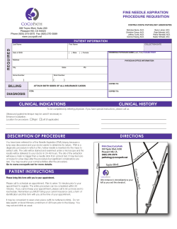
FIGURE 1. A 24-yr-old mentally disabled man presenting with fever and
rattling respiration had a normal chest radiography. The 111ln-lgG scan
shows abnormal activity in the right lung (posterior view). CT and pleural
puncture proved pleural empyema caused by S. pneumoniae. After antibiotic
therapy, fever and symptoms resolved (Patient 2).
U t i l i t y o f m 'In -Ig G in
FUO • de Kleijn et al.
487
TABLE 4
Diagnostic Utility of Scintigraphic Techniques in FUO in Literature
Investigators
No.
Scan
% helpful
scans
patients
Habibian et al. (75)
Hilson, Maisey (76)
Schmidt et al. (4 )
Syrjälä et al. (3)
Roddie et al. (5 )
Macsweeney et al. (77)
Davies et al. (78)
Kelly et al. (19)
Suga et al. (20)
Becker et al. (7)
Knockaert et al. (2)
Present study
22
67
32
68
17
25
28
28
36
34
145
58
67Ga
67Ga
111ln-oxine WBCs
111ln-oxine WBCs
99mTc-HMPAO WBCs
111ln-tropolonate WBCs
111ln-tropolonate WBCs
111ln-oxine WBCs
67Ga
""T c -a n ti NCA
67Ga
1111n-IgG
55
75
22
28
24
24
18
21
47
24
29
16
NCA = nonspecific cross-reacting antigen.
FIGURE 2. Cervical carcinoma was diagnosed in this 72-yr-old woman.
Surgery was unsuccessful and radiotherapy was administered. After 3 mo,
fever and abdominal pain developed. Abdominal US revealed a tumor
consistent with abnormal uptake in the lower abdomen on 1111n-IgG scintig
raphy. Laparotomy and culture revealed a pelvic abscess caused by Peptococcus spp. After surgery and antibiotic therapy, she recovered and her fever
resolved (Patient 5).
graphic imaging should not he a second step procedure in the
diagnostic work-up o f this subcategory o f patients with FUO.
CONCLUSION
During a 2-yr period, w^e prospectively investigated 167
patients with FUO. O f these patients, 58 underwent 111In-IgG
scintigraphy. These patients were prospectively separated in
patients with or without PDCs. Overall sensitivity and speci
ficity was 60% and 83%, respectively. In patients without PDCs
for local inflammation, the diagnostic yield o f scintigraphic
techniques is quite low since no focal inflammation was
observed. Therefore, 111 In-IgG scintigraphy should not be used
as a second-step procedure in the work-up o f these subgroup of
patients with FUO. In patients with PDCs at local inflammation,
m In-IgG is helpful in the diagnostic process in one-fourth of
the patients. This diagnostic yield is comparable with that o f the
ma jority of other scintigraphic techniques used in the diagnostic
process o f patients with FUO.
ACKNOWLEDGMENTS
We thank the members of The Netherlands FUO Study Group
for their contribution. This study was supported in part by The
Netherlands Institute for internal medicine through a grant from
Glaxo Inc. Zeist, The Netherlands and a grant from R.W. Johnson
Pharmaceutical Research Institute, Spring House, PA. Members of
the Netherlands FUO Imaging Group include: E.M.H.A. de Kleijn,
J.W.M. van der Meer, W.J.G. Oyen, F.H.M. Corstens, University
Hospital, St. Radboud, Nijmegen; H.G. Kreeftenberg and D.R.
Piers, University Hospital, Groningen; P. Speelman and E.A. van
Royen, University Hospital of the University of Amsterdam; S. de
Marie and E.P. Krenning, University Hospital Rotterdam.
REFERENCES
FIGURE 3. A 53-yr-old woman was referred from another hospital because
of fever of more than 3 wk duration. She also had a painful hip. Blood cultures
grew S. aureus and echocardiography revealed vegetations on the mitral
valve. The 1111n-IgG scintigraphy revealed metastatic abscesses in hip femur,
skull and chest. A culture of material obtained by puncture of the hip grew S.
aureus. After antibiotic therapy, the patient underwent cardiosurgery for valve
replacement. Thereafter, her fever disappeared (Patient 9).
488
T h e J o u r n a l o f N u c l e a r M edicine
1. Petersdorf RG, Beeson PB. Fever o f unexplained origin: report on 100 cases. Medicine
1961;40:1-30.
2. Knockaert DC, Mortelmans LA, de Roo MC, Bobbaers HJ. Clinical value o f
gallium-67 scintigraphy in evaluation o f fever o f unknown origin. Clin Infect Dis
1994;18:601-605.
3. Sytjülii MT. Valtonen V, Liewendahl K, Myllylä G. Diagnostic significance o f
in d iu m -1 11-granulocyte scintigraphy in febrile patients. J N u c l Med 1987;28:155-160.
4. Schmidt KG. Rasmussen JW, Sorrensen PG, W edcbye IM. Indium-111-granulocyte
scintigraphy in the evaluation o f patients with fever o f undetermined origin. Scand
./ Infect Dis 1987:19:339-345.
5. Roddie ME, Peters AM. Danpure HJ. et al. Inflammation: imaging with l,‘,mTcH M PA O -labeled leukocytes. Radiology 1988;166:767-772.
6. de Kleijn EM HA , Oyen WJG, Claesscn RAMJ. Corstens FHM, van der M eer JWM.
Utility o f scintigraphic methods in patients with fever o f unknown origin. Arch Intern
Med 1995:155:1989-1994.
7. Becker W. D ölkem eyer U, Gramatzki M. Schneider MU. Scheele J, W o lf F. Use o f
immunoscintigraphy in the diagnosis o f fever o f unknown origin. Eur ./ Nucl Med
1993;20:1078-1083.
8. Gardner P. Oster ZH. Rubor, calor. tumor and radionuclide scans. N Engl J Med
1989;321:970-972.
9. Knockaert DC. Vanneste LJ. Vanneste SB, Bobbaers 11J. Fever o f unknown origin in
the 1980s. An update o f the diagnostic spectrum. Arch Intern Med 1992; 15 2 : 5 1 55.
10. de Kleijn EM HA. van der Meer JW M. Fever o f unknown origin (FUO): report on 53
patients in a Dutch university hospital. Neth J Med 1995:47:54 60.
• Vol. 38 • No. 3 • March 1997
11. Larson EB. Feathcrstonc HJ, Petersdorf RG. Fever o f undetermined origin: diagnosis
and follow-up o f 105 cases, 1970-1980. Medicine 1982:61:269-292.
12. Palestro CJ. The current role o f gallium imaging in infection. Semin Nucl Med
1994;24:128-141.
13. Davis LP. Fink Bennett D. Nuclear medicine in the acutely ill patient. II. Crit Care
Clin 1994;10:383-400.
14. Serafim AN, Garty I, Vargas Cuba R, et al. Clinical evaluation o f a scintigraphic
method for diagnosing inflammations/infections using in d iu m -1 11 -labeled nonspecific
human IgG. J Nucl Med 1991;32:2227-2232.
15. Habibian MR, Staab EV, Mathews HA. Gallium-67 citrate scans in febrile patients.
JAMA 1975:233:1073-1076.
16. Hilson AJW . Maisey MN. Gallium-67 scanning in pyrexia o f unknown origin. Br
Med J 1979;279:1330-1331.
17. M acSwccney JE, Peters AM, Lavender JP. Indium-labeled leucocyte scanning in
pyrexia o f unknown origin. Clin Radiology 1 9 9 0 ;4 2 :4 1 4 -4 1 7 .
18. Davies SG, Garvie NW. The role o f indium-labeled leukocyte imaging in pyrexia o f
unknown origin. Br J Radiol 1990 ;63 :8 5 0 -8 5 4 .
19. Kelly MJ, K alff V, Hicks RJ. Spicer WJ, Spelman DW. Indium-111-oxine-labelcd
leukocyte scintigraphy in the detection and localization o f active inflammation and
sepsis. Med J Aus/ 1990;152:352-357.
20. Suga K, Nakagi K, Kuramitsu T, et al. The role o f 67Ga imaging in the detection o f foci
in recent cases o f fever o f unknown origin. Ann Nucl Med 1991;5:35-40.
Optimization of Technetium-99m-Labeled PEG
Liposomes to Image Focal Infection: Effects of
Particle Size and Circulation Time
Otto C. Boerman, Wim J.G. Oyen, Louis van Bloois, Emile B. Koenders, Jos W.M. van der Meer, Frans Fî.M. Corstens
and Gert Storni
Departments o f Nuclear Medicine and Internal Medicine, University>Hospital Nijmegen, Nijmegen; and Department o f
Pharmaceutics, Utrecht Institute for Pharmaceutical Sciences, Utrecht University, The Netherlands
r
In previous studies we have shown that liposomes sterically stabi
lized with polyethylene glycol (PEG), preferentially localize in infec
tious and inflammatory foci. In this study, we further optimized the
formulation of PEG liposomes for infection imaging in a rat model.
Methods: The biodistribution and imaging characteristics of differ
ent liposomal formulations labeled with 99mTc were determined in
rats with S. aureus infection of the left calf muscle. The influence of
liposomal size (mean diameter varying from 90 nm to 220 nm) as
well as circulation time (modulated by inclusion of 0-10 mole%
phosphatidylserine) were studied. Results: The smallest liposomes
displayed improved characteristics for infection imaging: 90-nm
liposomes revealed the highest abscess uptake (1.6% ± 0.4% ID/g,
24 hr postinjection) in combination with the lowest splenic accumu
lation (6.9% ± 0.7% ID/g, 24 hr postinjection) as compared to the
larger sized preparations. Enhanced abscess-to-blood ratios (4.0
versus 1.3 at 24 hr postinjection) were obtained by including 1.0
mole% phosphatidylserine in the lipid bilayer of the PEG liposomes.
However, enhanced blood clearance of these liposomes reduced
their absolute abscess uptake. Conclusion: These results indicate
that the in vivo behavior of PEG liposomes can be modulated to
optimize their characteristics for infection imaging.
Key Words: PEGylated liposomes; sterically stabilized liposomes;
S. aureus infection
J Nucl Med 1997; 38:489-493
L i p o s o m e s are microscopic lipid vesicles consisting o f one or
more concentric lipid bilayers enclosing discrete aqueous
spaces. Liposomes have been investigated extensively as carri
ers for drugs in attempts to achieve selective deposition and/or
controlled release of the encapsulated contents (7-5). In addi
tion, liposomes have been tested as vehicles to image infection
and inflammation (6,7). However, conventional liposomes are
rapidly taken up by cells o f the mononuclear phagocyte system
(MPS), which are primarily located in the liver and spleen (8,9).
A decade ago, one of the major goals in liposome research was
Received Apr. 16, 1996; revision accepted Jul. 3, 1996.
For correspondence or reprints contact: Otto C. Boerman, PhD, Dept, of Nuclear
Medicine, University Hospital Nijmegen, P.O. Box 9101, 6500 HB Nijmegen, The
Netherlands.
to enhance their circulatory residence time to allow enhanced
targeting to non-MPS tissues. It has been demonstrated that
small, neutral, cholesterol-rich liposomes composed o f rigid
phospholipids o f high-phase transition temperature show pro
longed circulation times at relatively high lipid doses (10-12).
More recently, it was demonstrated that inclusion o f polyethyleneglycol (PEG), conjugated to phosphatidylethanolamine in
the bilayer increased the blood circulation time as well (13,14).
This increment was at least as large as that observed with the
rigid lipid composition but without the requirements o f specific
lipid composition, particle size and lipid dose (15-17). The
prolonged circulation time of PEG liposomes, also referred to as
sterically stabilized or StealthK liposomes (Sequus Pharmaceu
ticals Inc., Menlo Park, CA), is caused by reduced recognition
by the MPS, as reflected by delayed and diminished hepatic and
splenic accumulation. The development o f long-circulating
liposomal formulations has offered several new applications for
liposomes such as; (a) long-term controlled release o f drugs in
the circulation; (b) improved antibody-guided delivery o f lipo
somes; and (c) enhanced targeting to non-MPS-related patho
logical sites such as tumors and inflammatory foci (18,19).
Our previous studies in rats have shown that PEG liposomes
labeled with either 11'in or " mTc may be excellent radiophar
maceuticals for imaging infectious and inflammatory foci (1,2).
The aim o f this study was to tailor the PEG-liposomal formu
lation for scintigraphic application in rats with focal S. aureus
infection. The PEG-liposomal formulation we used in our
previous studies was originally developed for controlled deliv
ery o f chemotherapeutics (15,20,21). In this study, we modified
the size and lipid composition o f the liposomes to optimize their
in vivo behavior for imaging infection. Different liposome
dispersions with a narrow size distribution were produced
(mean size: 90, 120, 160 and 220 nm) and evaluated in vivo. In
addition, the effects o f enhanced blood clearance were investi
gated by incorporating increasing amounts o f phosphatidylser
ine (PS) (0, 1 and 10 mole%) in the lipid bilayer. It has been
shown that PS exposure strongly increases the recognition o f
PEG liposomes by macrophages, thereby causing enhanced
blood clearance (22,23).
PEG
L iposom es f o r I n f e c tio n Im ag in g
• Boerman et al.
489
© Copyright 2026 ExpyDoc